. 2013;15(6):R219.
doi: 10.1186/ar4416.

Deficient spontaneous in vitro apoptosis and increased tmTNF reverse signaling-induced apoptosis of monocytes predict suboptimal therapeutic response of rheumatoid arthritis to TNF inhibition

Deficient spontaneous in vitro apoptosis and increased tmTNF reverse signaling-induced apoptosis of monocytes predict suboptimal therapeutic response of rheumatoid arthritis to TNF inhibition

Undine Meusch et al. Arthritis Res Ther. 2013.

Abstract

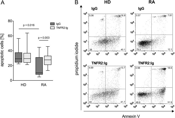
Introduction: In vitro apoptosis of peripheral monocytes in rheumatoid arthritis (RA) is disturbed and influenced by cytokine production and transmembrane TNF (tmTNF) reverse signaling. The goal of the study was the analysis of the predictive value of the rate of in vitro apoptosis for the therapeutic response to anti-TNF treatment.

Methods: Spontaneous and tmTNF reverse signaling-induced apoptosis were determined in vitro in monocytes from 20 RA patients prior to initiation of therapeutic TNF inhibition with etanercept, and the subsequent clinical response was monitored.

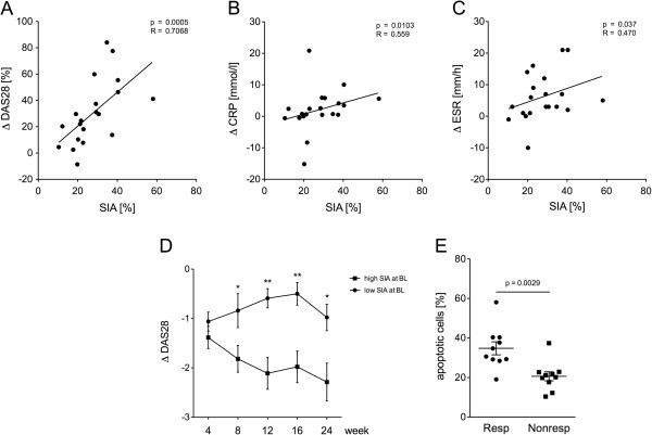
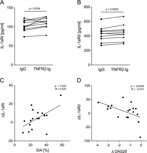
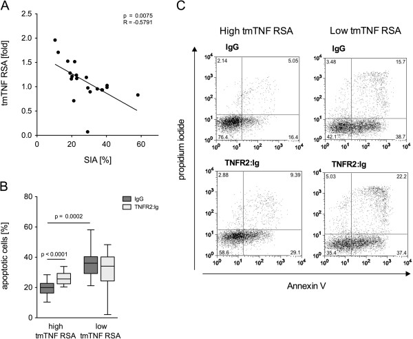
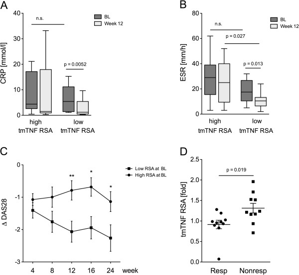
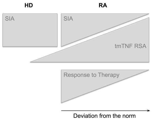
Results: Spontaneous in vitro apoptosis was significantly reduced in RA patients compared to controls. Deficiency in spontaneous apoptosis was associated with an insufficient therapeutic response according to the European League Against Rheumatism (EULAR) response criteria and less reduction of the disease activity determined by disease activity score (DAS) 28. High susceptibility to reverse signaling-induced apoptosis was also associated with less efficient reduction in the DAS28. Of note, a strong negative correlation between the two apoptotic parameters was discernible, possibly indicative of two pathogenetically relevant processes counter-regulating each other. tmTNF reverse signaling induced in vitro production of soluble IL1-RI and IL-1RII only in monocytes not deficient in spontaneous apoptosis, and the levels of soluble IL1-RII were found to be predictive of a good clinical response to Etanercept.

Conclusion: Although tmTNF reverse signaling is able to induce apoptosis of RA monocytes in vitro, this process appears to occur in vitro preferentially in patients with suboptimal therapeutic response. Resistance to spontaneous in vitro apoptosis, in contrast, is a predictor of insufficient response to treatment.


Figures

Figure 1
Figure 1
TNFR2:Ig triggered reverse signaling via transmembrane (tm)TNF induces apoptosis in monocytes from rheumatoid arthritis (RA) patients but not from healthy controls. (A) Monocytes from the peripheral blood of healthy donors (HD) and from RA patients (RA) were cultured with IgG or TNFR2:Ig for 16 hours. Box plot depicts the rate of spontaneous apoptosis in vitro (SIA) cultures with IgG (dark gray bars) and of tmTNF reverse signaling-induced apoptosis (tmTNF RSA) in the presence of TNFR2:Ig (light gray bars) in healthy controls (HD) (n = 10) and RA patients (RA) (n = 13). Significant differences are as indicated. (B) Representative dot plots of annexin V- and propidium iodide-stained monocytes from a healthy control (left panels) and from one RA patient (right panels) after incubation with IgG or TNFR2:Ig for 16 hours. Upper panels show spontaneous apoptosis in vitro (SIA) (IgG), lower panels tmTNF reverse signaling induced apoptosis (tmTNF RSA) (TNFR2:Ig).
Figure 2
Figure 2
Spontaneous in vitro apoptosis of monocytes correlates with change in disease activity after 12 weeks. (A-C) Spontaneous apoptosis of monocytes from rheumatoid arthritis (RA) patients was measured at baseline and the change in disease activity score (DAS)28, C-reactive protein (CRP) and erythrocyte sedimentation rate (ESR) levels from baseline and to week 12 after starting treatment with etanercept for these patients was calculated. Dot plots depict the rate of spontaneous in vitro apoptosis (SIA) in relation to the change (Δ) in DAS28 (A), CRP (B) and ESR (C) (n = 20). Regression coefficient and level of significance for the linear regression are as indicated. (D) Processing of DAS28 during the study was analyzed in the patient cohorts with high and low spontaneous monocyte apoptosis respectively. Change in DAS28 was calculated for the treatment time points 4, 8 12, 16 and 24 weeks. Depicted are median and standard error of the mean of numeric improvement in DAS28 (ΔDAS28) in the patient group with high (solid squares) and low (solid circles) rates of spontaneous in vitro SIA at baseline (BL) at indicated time points (n = 20). For significant differences between ΔDAS28 in both groups at different time points, significance levels are indicted: *P <0.05, **P <0.01. (E) Dot plot depicts the rate of spontaneous in vitro SIA in patients achieving a good therapeutic response after 12 weeks of etanercept (responder, Resp) (n = 20) and in patients with only moderate or no response (nonresponders, Nonresp). Significant differences are as indicated.
Figure 3
Figure 3
Transmembrane (tm)TNF reverse signaling (RS)-induced secretion of soluble IL1-RI and IL1-RII depends on apoptotic rates and influences clinical response. (A, B) tmTNF RS-induced IL-1RI and IL-1RII expression was measured in in vitro monocyte cultures from rheumatoid arthritis (RA) patients after baseline incubation IgG and TNFR2:Ig for 16 hours. Individually paired data of IL1sRI (A) and IL1sRII (B) concentrations in control cultures (IgG) (solid circles) and in cultures with TNFR2:Ig (solid triangles) determined in vitro at baseline in monocytes from the subgroup of RA patients (n = 10) characterized by high rates of spontaneous in vitro apoptosis (SIA) are as indicated. Significant differences are indicated. (C) Levels of tmTNF RS-induced IL-1RI production (ΔIL-1sRI) by RA monocytes at baseline correlates with spontaneous monocyte apoptosis in these RA patients. Dot plots depicts rate of SIA in relation to the baseline concentration of tmTNF RS-induced IL1sRI (n = 18). ∆IL-1sRI is the difference of tmTNF RS-induced IL-1sRI production and spontaneous IL-1sRI production. Regression coefficient and significane level for linear regression are indicated. (D) Levels of baseline tmTNF RS-induced IL-1RII production by RA monocytes correlated negatively with change (Δ) in disease activity score (DAS)28 12 weeks after starting treatment. Dot plots depicts numeric improvement in (ΔDAS28) in RA patients (n = 18) after 12 weeks of TNF blockade in relation to the concentration of tmTNF RS-induced IL1-sRII (ΔIL-1sRII) at baseline. ∆IL-1sRII is the difference in tmTNF RS-induced IL-1sRII production and spontaneous IL-1sRII production. Regression coefficient and level of significance for the linear regression are as indicated.
Figure 4
Figure 4
Monocytes with low spontaneous in vitro apoptosis are susceptible for transmembrane TNF reverse signaling-induced apoptosis (tmTNF RSA). (A) Dot plot shows the rate of spontaneous in vitro apoptosis (SIA) in relation to tmTNF RSA) in rheumatoid arthritis (RA) patients (n = 20). tmTNF RSA is shown as ratio of TNFR2:Ig apoptosis and SIA. Regression coefficient and level of significance for the linear regression are as indicated. (B) Box plot depicts the rate of tmTNF RSA (light gray bars, TNFR2:Ig) in comparison to SIA (dark gray bars, IgG) in the groups with high and low tmTNF RSA separately (n = 20). Significant differences are as indicated. (C) Representative dot plots of annexin V- and propidium iodide-stained monocytes from one patient with high tmTNF RSA (left panels) and one with low TNF RSA (right panels). Upper panels show SIA (IgG), lower panels TNF RSA (TNFR2:Ig).
Figure 5
Figure 5
Transmembrane (tm)TNF reverse signaling induced apoptosis of monocytes correlates with change in disease activity after 12 weeks. (A, B) Box plots depict C-reactive protein (CRP) (A) and erythrocyte sedimentation rate (ESR) (B) at baseline (BL) (dark gray bars) and after 12 weeks of etanercept (light gray bars) in the groups with high tmTNF reverse signaling induced apoptosis (tmTNF RSA) and low tm tmTNF RSA separately (n = 20). Significant differences are as indicated. (C) Depicted are median and standard error of the mean of the numeric improvement (Δ) in disease activity score (DAS)28 in the patient group with high (solid squares, n = 11) and low (solid circles, n = 9) rates of tmTNF RSA at the indicated time points. For significant differences between ΔDAS28 in both groups at the different time points, the levels of significance are indicated: *P <0.05, **P <0.01). (D) Dot plot depicts the rate of tmTNF RSA in patients achieving a good therapeutic response after 12 weeks of etanercept (responder, Resp) and in patients with only a moderate or no response (nonresponders, Nonresp). Significant differences are as indicated.
Figure 6
Figure 6
Schematic representation of the magnitude of spontaneous in vitro apoptosis (SIA) and transmembrane TNF reverse signaling-induced apoptosis (tmTNF RSA) in healthy donors (HD) and patients with rheumatoid arthritis (RA). A shift from SIA towards tmTNF RSA is associated with both disease severity and the clinical response under anti-TNF treatment.

Similar articles

Cited by

References

    1. Mitoma H, Horiuchi T, Hatta N, Tsukamoto H, Harashima SI, Kikuchi Y, Otsuka J, Okamura S, Fujita S, Harada M. Infliximab induces potent anti-inflammatory responses by outside-to-inside signals through transmembrane TNF-alpha. Gastroenterology. 2005;15:376–392. doi: 10.1053/j.gastro.2004.11.060. - DOI - PubMed
    1. Mitoma H, Horiuchi T, Tsukamoto H, Tamimoto Y, Kimoto Y, Uchino A, To K, Harashima SI, Hatta H, Harada M. Mechanisms for cytotoxic effects of anti-tumor necrosis factor agents on transmembrane tumor necrosis factor alpha-expressing cells: comparison among infliximab, etanercept, and adalimumab. Arthritis Rheum. 2008;15:1248–1257. doi: 10.1002/art.23447. - DOI - PubMed
    1. Rossol M, Schubert K, Meusch U, Schulz A, Biedermann B, Grosche J, Pierer M, Scholz R, Baerwald C, Thiel A, Hagen S, Wagner U. Tumor necrosis factor receptor type I expression of CD4+ T cells in rheumatoid arthritis enables them to follow tumor necrosis factor gradients into the rheumatoid synovium. Arthritis Rheum. 2013;15:1468–1476. doi: 10.1002/art.37927. - DOI - PubMed
    1. Makrygiannakis D, Catrina AI. Apoptosis as a mechanism of action of tumor necrosis factor antagonists in rheumatoid arthritis. J Rheumatol. 2012;15:679–685. doi: 10.3899/jrheum.110974. - DOI - PubMed
    1. Meusch U, Rossol M, Baerwald C, Hauschildt S, Wagner U. Outside-to-inside signaling through transmembrane tumor necrosis factor reverses pathologic interleukin-1beta production and deficient apoptosis of rheumatoid arthritis monocytes. Arthritis Rheum. 2009;15:2612–2621. doi: 10.1002/art.24778. - DOI - PubMed

Publication types

MeSH terms