Vertebral artery position in the setting of cervical degenerative disease: implications for selective cervical transforaminal epidural injections
- PMID: 24355145
- PMCID: PMC3902740
- DOI: 10.1177/159101991301900404
Vertebral artery position in the setting of cervical degenerative disease: implications for selective cervical transforaminal epidural injections
Abstract
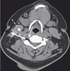
Cervical transforaminal epidural injections (C-TfEI) are commonly performed in patients with cervical radiculopathy/pain. C-TfEIs are typically performed without incident but adverse events can occur. Using CT-fluoroscopy-guided C-TfEI, we commonly observe the vertebral artery in proximity to the target injection site. The purpose of this study was to assess the position of the vertebral artery relative to the typical C-TfEI injection point. CT-fluoroscopy-guided C-TfEIs were performed at 70 levels in 68 patients with radiculopathy/neck pain (age range 19-83 yrs, mean 50.6 yrs). Degenerative neural foraminal narrowing at each level was characterized (normal-to-mild, moderate, severe). Vertebral artery position was categorized as: anterior (normal), partially covering neural foramen, complete/near-complete covering the neural foramen. Additional measured variables included angle of needle trajectory, foraminal angle, and whether or not needle trajectory intersected with the vertebral artery. Foraminal vertebral artery covering correlated with severity of foraminal degenerative narrowing (p=0.003). Complete/near-complete covering was seen in: 65% severely narrowed foramina, 30% moderately narrowed foramina and 10% normal/mildly-narrowed foramina. Needle trajectory intersected with the vertebral artery in 30 of 70 injections (46%) by CT-fluoroscopy, frequently associated with shallow (lateral) approaches. Foraminal angle, approximating oblique fluoroscopic technique, suggests needle trajectory intersection with the vertebral artery in 27 of 70 foramina (39%). Vertebral artery position is commonly displaced into the foramen in patients with advanced cervical degenerative disease. Operator awareness of altered vertebral artery position is important for determination of optimal needle trajectory and tip placement prior to injection in patients undergoing C-TfEI.
Keywords: cervical nerve root block; cervical radiculopathy; cervical spine; cervical transforaminal epidural injection; computed tomography; neck pain.
Figures


Similar articles
-
New Optimal Needle Entry Angle for Cervical Transforaminal Epidural Steroid Injections: A Retrospective Study.Int J Med Sci. 2017 Apr 8;14(4):376-381. doi: 10.7150/ijms.17112. eCollection 2017. Int J Med Sci. 2017. PMID: 28553170 Free PMC article.
-
Novel Method for S1 Transforaminal Epidural Steroid Injection.World Neurosurg. 2020 Jan;133:e443-e447. doi: 10.1016/j.wneu.2019.09.051. Epub 2019 Sep 14. World Neurosurg. 2020. PMID: 31526885
-
A Prospective Comparison of CT-Epidurogram Between Th1-Transforaminal Epidural Injection and Th1/2-Parasagittal Interlaminar Epidural Injection for Cervical Upper Limb Pain.Pain Physician. 2019 Mar;22(2):165-176. Pain Physician. 2019. PMID: 30921982 Clinical Trial.
-
Cervical radicular pain: the role of interlaminar and transforaminal epidural injections.Curr Pain Headache Rep. 2014 Jan;18(1):389. doi: 10.1007/s11916-013-0389-9. Curr Pain Headache Rep. 2014. PMID: 24338702 Review.
-
Adverse central nervous system sequelae after selective transforaminal block: the role of corticosteroids.Spine J. 2004 Jul-Aug;4(4):468-74. doi: 10.1016/j.spinee.2003.10.007. Spine J. 2004. PMID: 15246308 Review.
Cited by
-
Neck movement during cervical transforaminal epidural injections and the position of the vertebral artery: an anatomical study.Acta Radiol Open. 2019 Mar 12;8(3):2058460119834688. doi: 10.1177/2058460119834688. eCollection 2019 Mar. Acta Radiol Open. 2019. PMID: 30886742 Free PMC article.
-
An open-label non-inferiority randomized trail comparing the effectiveness and safety of ultrasound-guided selective cervical nerve root block and fluoroscopy-guided cervical transforaminal epidural block for cervical radiculopathy.Ann Med. 2022 Dec;54(1):2681-2691. doi: 10.1080/07853890.2022.2124445. Ann Med. 2022. PMID: 36164681 Free PMC article. Clinical Trial.
-
Cervical Selective Nerve Root Block: Three-dimensional Puncture Planning With Dyna-CT Is Superior to Conventional CT-guidance in an Ex Vivo Model.In Vivo. 2025 Mar-Apr;39(2):713-723. doi: 10.21873/invivo.13875. In Vivo. 2025. PMID: 40010983 Free PMC article.
-
Combined fluoroscopic and ultrasound guided cervical nerve root injections.Int Orthop. 2016 Dec;40(12):2547-2551. doi: 10.1007/s00264-016-3224-1. Epub 2016 May 24. Int Orthop. 2016. PMID: 27222157
-
Cervical foraminal steroid injections under CT guidance: retrospective study of in situ contrast aspects in a serial of 248 cases.Skeletal Radiol. 2015 Jan;44(1):1-8. doi: 10.1007/s00256-014-2028-x. Epub 2014 Oct 16. Skeletal Radiol. 2015. PMID: 25316168 Review.
References
-
- Anderberg L, Säveland H, Annertz M. Distribution patterns of transforaminal injections in the cervical spine evaluated by multi-slice computed tomography. Eur Spine J. 2006;15(10):1465–1471. - PubMed
-
- Hoang JK, Apostol MA, Kranz PG, et al. CT fluoroscopy-assisted cervical transforaminal steroid injection: tips, traps, and use of contrast material. Am J Roentgenol. 2010;195(4):888–894. - PubMed
-
- Vallée JN, Feydy A, Carlier RY, et al. Chronic cervical radiculopathy: lateral-approach periradicular corticosteroid Injection. Radiology. 2001;218(3):886–892. - PubMed
Publication types
MeSH terms
Substances
LinkOut - more resources
Full Text Sources
Other Literature Sources

