Case Reports
. 2013 Dec;19(4):483-8.
doi: 10.1177/159101991301900413. Epub 2013 Dec 18.

Bilateral cervical spinal dural arteriovenous fistulas with intracranial venous drainage mimicking a foramen magnum dural arteriovenous fistula

Affiliations
Case Reports

Bilateral cervical spinal dural arteriovenous fistulas with intracranial venous drainage mimicking a foramen magnum dural arteriovenous fistula

Steven W Hetts et al. Interv Neuroradiol. 2013 Dec.

Abstract

We describe a unique case of bilateral cervical spinal dural arteriovenous fistulas mimicking an intracranial dural arteriovenous fistula near the foramen magnum. We review its detection via MRI and digital subtraction angiography and subsequent management through surgical intervention. Pitfalls in diagnostic angiography are discussed with reference to accurate location of the fistula site. The venous anastomotic connections of the posterior midline spinal vein to the medial posterior medullary vein, posterior fossa bridging veins, and dural venous sinuses of the skull base are discussed with reference to problem-solving in this complex case. The mechanism of myelopathy through venous hypertension produced by spinal dural fistulas is also emphasized.

Keywords: MR angiography; MR imaging; digital subtraction angiography; spinal angiography; spinal dural arteriovenous fistula; spine; vascular malformations.


Figures
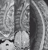
Figure 1
Figure 1
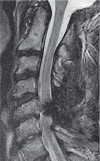
Sagittal and axial T2 weighted MR images of the cervical and thoracic spine at the time of initial presentation demonstrate patchy discontinuous long-segment T2 prolongation in the central cervical and thoracic spinal cord (A-D) with associated enlarged flow voids along the dorsal surface of the cervical spinal cord and cervicomedullary junction (A,B). Degenerative spinal canal stenosis is also evident in the lower cervical spine, severe at C5/6. Axial FLAIR images of the brain demonstrate additional focal T2 hyperintense white matter lesions involving the corticospinal tracts (E) and brachium pontis (F).
Figure 2
Figure 2
Sagittal T2 MRI 5 months after initial presentation shows resolution of spinal cord edema, but persistence of enlarged flow voids dorsal to the cervical spinal cord and medulla (A). DSA shows right (D,E) and left (F) costocervical artery supply to the enlarged midline intrathecal vessel, confirmed to run dorsal to the spinal cord by injection of the right costocervical artery during lateral DSA (B). Dural entry point of the shunting vessel (*) and presumed fistula site at the exit point to the epidural venous plexus (black arrows) are marked.
Figure 3
Figure 3
Frontal (A) and lateral (D) DSA of the right costocervical artery following surgical clipping of the intradural vessel in the posterior fossa at its entry point into the dura (*) as well as its exit point to the epidural venous plexus (black arrow). Although there is no longer filling of the epidural venous plexus between the occiput and C1, there has been redirection of blood flow to veins along the surface of the medulla, pons, and midbrain (white arrows) that now decompress into the cavernous sinuses (cs) bilaterally. Similar results were found upon injection of the left costocervical artery (not shown).
Figure 4
Figure 4
Sagittal T2 weighted MRI of the cervical spine performed 6 months postoperatively shows no persistent or recurrent dilated intrathecal flow voids and no T2 prolongation in the spinal cord (as limited at C5/6 by metallic artifact from surgical clips). A brain MRI performed at the same time (not shown) also demonstrated resolution of white matter lesions in the posterior fossa and corticospinal tracts.

References

    1. Krings T, Geibprasert S. Spinal dural arteriovenous fistulas. Am J Neuroradiol. 2009;30:639–648. - PMC - PubMed
    1. Narvid J, Hetts SW, Larsen D, et al. Spinal dural arteriovenous fistulae: clinical features and long-term results. Neurosurgery. 2008;62:159–166. discussion 166-157. - PubMed
    1. Brunereau L, Gobin YP, Meder JF, et al. Intracranial dural arteriovenous fistulas with spinal venous drainage: relation between clinical presentation and angiographic findings. Am J Neuroradiol. 1996;17:1549–1554. - PMC - PubMed
    1. Hetts SW, Moftakhar P, English JD, et al. Spinal dural arteriovenous fistulas and intrathecal venous drainage: correlation between digital subtraction angiography, magnetic resonance imaging, and clinical findings. J Neurosurg Spine. 2012;16:433–440. - PubMed
    1. Oshita J, Yamaguchi S, Ohba S, et al. Mirror-image spinal dural arteriovenous fistulas at the craniocervical junction: case report and review of the literature. Neurosurgery. 2011;69:E1166–1171. - PubMed

Publication types