

Lhermitte-Duclos disease. A case report
- PMID: 24355184
- PMCID: PMC4202883
- DOI: 10.1177/197140091302600608
Lhermitte-Duclos disease. A case report
Abstract
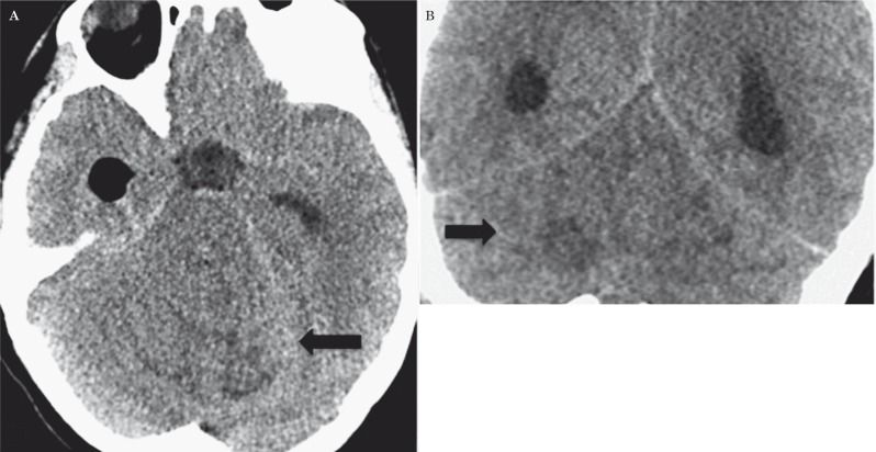
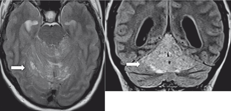
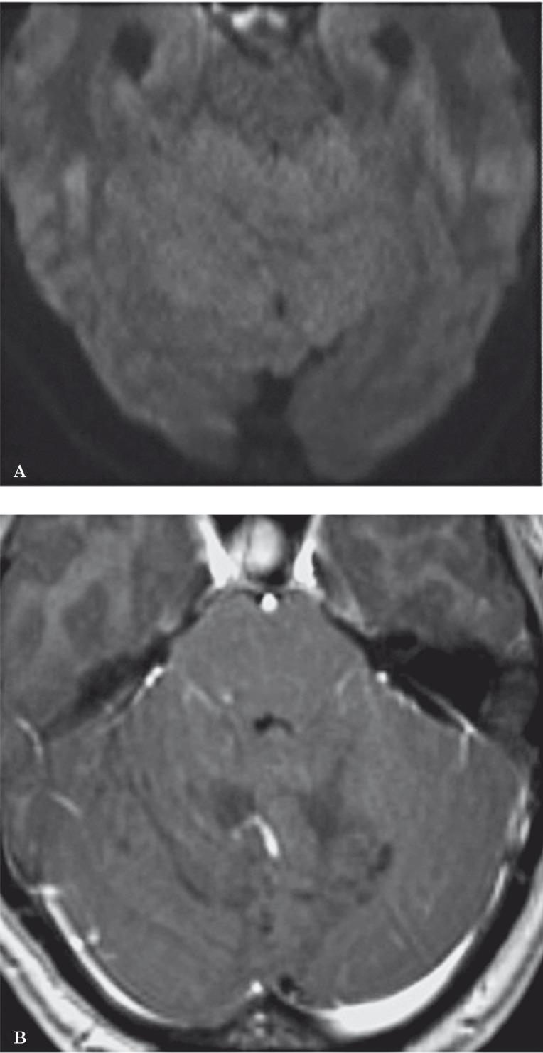
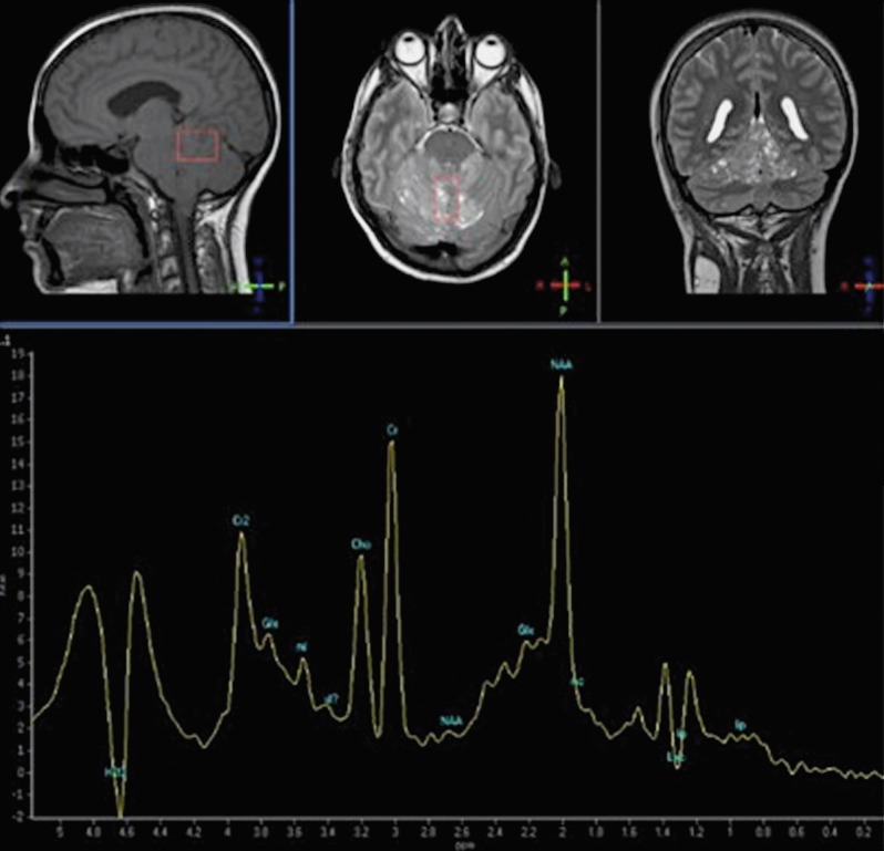
Lhermitte-Duclos disease is a rare pathologic condition consisting of a dysplastic gangliocytoma of the cerebellum. Its association with phacomatosis and an autosomal dominant neoplastic syndrome, Cowden's syndrome is also known. Modern neuroimaging contributes to a correct diagnosis and pre- and postoperative evaluation. Here we describe the morphologic and metabolic aspects of the disease as shown by conventional MRI, diffusion imaging and spectroscopy in a 31-year-old woman. In addition, the specific neuroradiologic characteristics are presented and discussed in the light of the main pathologic and clinical features, such as hypertrophy of the cerebellar folia associated with white matter atrophy.
Keywords: CT and MR imaging; Lhermitte-Duclos disease; brain tumor.
Figures
References
-
- Nowak DA, Trost HA. Lhermitte-Duclos disease (dysplastic cerebellar gangliocytoma): a malformation, hamartoma or neoplasm? Acta Neurol Scand. 2002;105(3):137–145. - PubMed
-
- Kulkantrakorn K, Awwad EE, Levy E, et al. MRI in Lhermitte-Duclos disease. Neurology. 1997;48(3):725–731. - PubMed
-
- Murray C, Shipman P, Khangure M, et al. Lhermitte-Duclos disease associated with Cowden’s syndrome: case report and literature review. Australas Radiol. 2001;45(3):343–346. - PubMed
-
- Miller C. Cowden disease (multiple hamartoma syndrome) In: Elmets C, Vinson R, Libow L, Quirk C, James W W, et al., editors. EMEDICINE instant access to the minds of medicine. LU; 2002.
Publication types
MeSH terms
LinkOut - more resources
Full Text Sources
Other Literature Sources
