

REEP1 and REEP2 proteins are preferentially expressed in neuronal and neuronal-like exocytotic tissues
- PMID: 24355597
- PMCID: PMC3919455
- DOI: 10.1016/j.brainres.2013.12.008
REEP1 and REEP2 proteins are preferentially expressed in neuronal and neuronal-like exocytotic tissues
Abstract
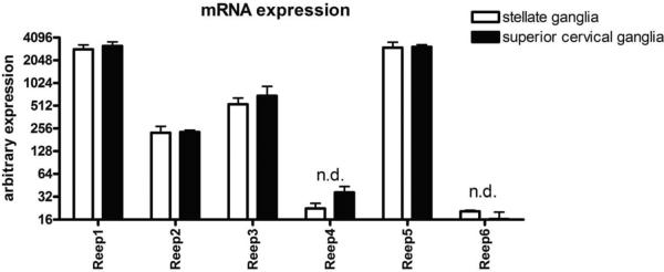
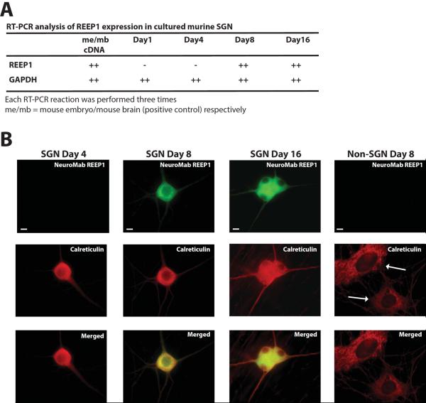
The six members of the Receptor Expression Enhancing Protein (REEP) family were originally identified based on their ability to enhance heterologous expression of olfactory receptors and other difficult to express G protein-coupled receptors. Interestingly, REEP1 mutations have been linked to neurodegenerative disorders of upper and lower motor neurons, hereditary spastic paraplegia (HSP) and distal hereditary motor neuropathy type V (dHMN-V). The closely related REEP2 isoform has not demonstrated any such disease linkage. Previous research has suggested that REEP1 mRNA is ubiquitously expressed in brain, muscle, endocrine, and multiple other organs, inconsistent with the neurodegenerative phenotype observed in HSP and dHMN-V. To more fully examine REEP1 expression, we developed and characterized a new REEP1 monoclonal antibody for both immunoblotting and immunofluorescent microscopic analysis. Unlike previous RT-PCR studies, immunoblotting demonstrated that REEP1 protein was not ubiquitous; its expression was restricted to neuronal tissues (brain, spinal cord) and testes. Gene expression microarray analysis demonstrated REEP1 and REEP2 mRNA expression in superior cervical and stellate sympathetic ganglia tissue. Furthermore, expression of endogenous REEP1 was confirmed in cultured murine sympathetic ganglion neurons by RT-PCR and immunofluorescent staining, with expression occurring between Day 4 and Day 8 of culture. Lastly, we demonstrated that REEP2 protein expression was also restricted to neuronal tissues (brain and spinal cord) and tissues that exhibit neuronal-like exocytosis (testes, pituitary, and adrenal gland). In addition to sensory tissues, expression of the REEP1/REEP2 subfamily appears to be restricted to neuronal and neuronal-like exocytotic tissues, consistent with neuronally restricted symptoms of REEP1 genetic disorders.
Keywords: AR; Distal hereditary motor neuropathy type V; G protein-coupled receptor; GPCR; HSP; Hereditary spastic paraplegia; NGF; Neurodegeneration; OR; REEP; RT-PCR; RTP; Receptor expression enhancing protein; SCG; SG; Sympathetic ganglion neuron; Yip; Ypt interacting protein; adrenergic receptor; dHMN-V; distal hereditary motor neuropathy type V; hereditary spastic paraplegia; mAb; monoclonal antibody; nerve growth factor; olfactory receptor; receptor expression enhancing protein; receptor transporting protein; reverse transcription polymerase chain reaction; stellate ganglion, SGN, sympathetic ganglion neurons; superior cervical ganglia.
Copyright © 2013 Elsevier B.V. All rights reserved.
Figures


Similar articles
-
REEPs are membrane shaping adapter proteins that modulate specific g protein-coupled receptor trafficking by affecting ER cargo capacity.PLoS One. 2013 Oct 2;8(10):e76366. doi: 10.1371/journal.pone.0076366. eCollection 2013. PLoS One. 2013. PMID: 24098485 Free PMC article.
-
Exome sequencing identifies a REEP1 mutation involved in distal hereditary motor neuropathy type V.Am J Hum Genet. 2012 Jul 13;91(1):139-45. doi: 10.1016/j.ajhg.2012.05.007. Epub 2012 Jun 14. Am J Hum Genet. 2012. PMID: 22703882 Free PMC article.
-
A spastic paraplegia mouse model reveals REEP1-dependent ER shaping.J Clin Invest. 2013 Oct;123(10):4273-82. doi: 10.1172/JCI65665. Epub 2013 Sep 24. J Clin Invest. 2013. PMID: 24051375 Free PMC article.
-
The REEP family of proteins: Molecular targets and role in pathophysiology.Pharmacol Res. 2022 Nov;185:106477. doi: 10.1016/j.phrs.2022.106477. Epub 2022 Sep 30. Pharmacol Res. 2022. PMID: 36191880 Review.
-
A complete overview of REEP1: old and new insights on its role in hereditary spastic paraplegia and neurodegeneration.Rev Neurosci. 2020 May 26;31(4):351-362. doi: 10.1515/revneuro-2019-0083. Rev Neurosci. 2020. PMID: 31913854 Review.
Cited by
-
The Mycobacterium tuberculosis H37Ra gene MRA_1916 causes growth defects upon down-regulation.Sci Rep. 2015 Nov 4;5:16131. doi: 10.1038/srep16131. Sci Rep. 2015. PMID: 26531045 Free PMC article.
-
Protocol for using fluorescent sensors targeted to endogenous proteins (FluoSTEPs) to measure microdomain-specific signaling events.STAR Protoc. 2021 Aug 2;2(3):100693. doi: 10.1016/j.xpro.2021.100693. eCollection 2021 Sep 17. STAR Protoc. 2021. PMID: 34467224 Free PMC article.
-
Hereditary spastic paraplegia-linked REEP1 modulates endoplasmic reticulum/mitochondria contacts.Ann Neurol. 2015 Nov;78(5):679-96. doi: 10.1002/ana.24488. Epub 2015 Sep 16. Ann Neurol. 2015. PMID: 26201691 Free PMC article.
-
Aurora kinase A-mediated phosphorylation triggers structural alteration of Rab1A to enhance ER complexity during mitosis.Nat Struct Mol Biol. 2024 Feb;31(2):219-231. doi: 10.1038/s41594-023-01165-7. Epub 2024 Jan 4. Nat Struct Mol Biol. 2024. PMID: 38177680
-
REEP1 Preserves Motor Function in SOD1G93A Mice by Improving Mitochondrial Function via Interaction with NDUFA4.Neurosci Bull. 2023 Jun;39(6):929-946. doi: 10.1007/s12264-022-00995-7. Epub 2022 Dec 15. Neurosci Bull. 2023. PMID: 36520405 Free PMC article.
References
-
- Bartlett JM. Approaches to the analysis of gene expression using mRNA: a technical overview. Mol Biotechnol. 2002;21:149–60. - PubMed
Publication types
MeSH terms
Substances
Grants and funding
LinkOut - more resources
Full Text Sources
Other Literature Sources
Medical
Research Materials
