Review
. 2013 Dec 16;23(24):R1113-21.
doi: 10.1016/j.cub.2013.11.009.

Nuclear mechanics and mechanotransduction in health and disease

Affiliations
Review

Nuclear mechanics and mechanotransduction in health and disease

Philipp Isermann et al. Curr Biol. .

Abstract

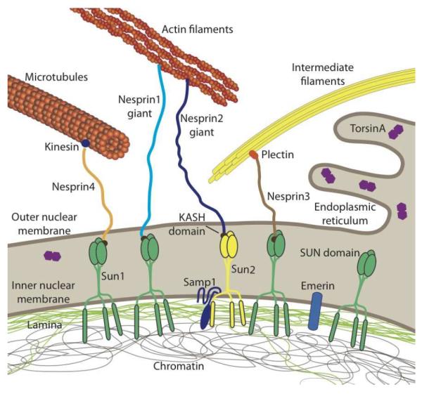
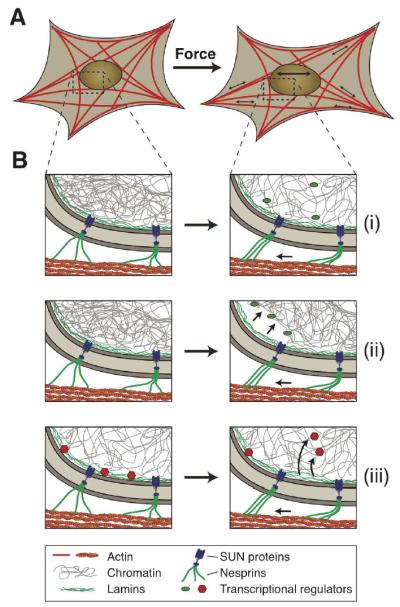
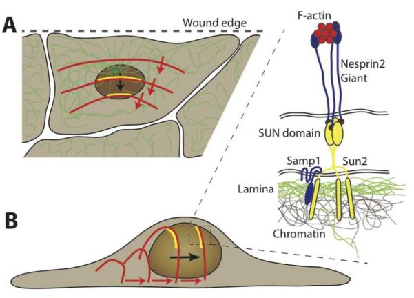
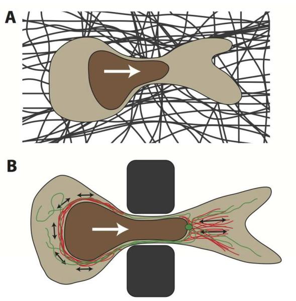
The nucleus is the defining feature of eukaryotic cells and often represents the largest organelle. Over the past decade, it has become apparent that the nucleus is tightly integrated into the structural network of the cell through so-called LINC (linker of the nucleoskeleton and cytoskeleton) complexes, which enable transmission of forces between the nucleus and cytoskeleton. This physical connection between the nucleus and the cytoskeleton is essential for a broad range of cellular functions, including intracellular nuclear movement and positioning, cytoskeletal organization, cell polarization, and cell migration. Recent reports further indicate that forces transmitted from the extracellular matrix to the nucleus via the cytoskeleton may also directly contribute to the cell's ability to probe its mechanical environment by triggering force-induced changes in nuclear structures. In addition, it is now emerging that the physical properties of the nucleus play a crucial role during cell migration in three-dimensional (3D) environments, where cells often have to transit through narrow constrictions that are smaller than the nuclear diameter, e.g., during development, wound healing, or cancer metastasis. In this review, we provide a brief overview of how LINC complex proteins and lamins facilitate nucleo-cytoskeletal coupling, highlight recent findings regarding the role of the nucleus in cellular mechanotransduction and cell motility in 3D environments, and discuss how mutations and/or changes in the expression of these nuclear envelope proteins can result in a broad range of human diseases, including muscular dystrophy, dilated cardiomyopathy, and premature aging.


Figures

Figure 1
Figure 1. Schematic overview of LINC complex proteins and their connections to the cytoskeleton and nuclear interior
SUN proteins at the inner nuclear membrane bind to the nuclear lamina and other nucleoplasmic proteins while interacting with KASH-domain containing proteins at the outer nuclear membrane. KASH-domain containing proteins directly or indirectly interact with cytoskeletal filaments, thereby forming a physical connection between the nuclear interior and cytoskeleton. Please note that SUN- and KASH domain proteins can exist in multiple isoforms encoded by several genes. In human somatic cells, the most predominant KASH-domain proteins are nesprin-1, -2, and -3 and their various isoforms, and Sun1 and Sun2 as the predominant SUN proteins [16]. Illustrated are only the largest isoforms for nesprins1–4; cells express many additional shorter nesprin isoforms, including some lacking the KASH domain. Smaller nesprin isoform may also be located on the inner nuclear membrane. Note that nesprin-1, -2, -4 and KASH5 can also interact with kinesin and/or dynein. Samp1 and torsinA are involved in the regulation of the LINC complex. Not depicted are KASH5 and the SUN protein isoforms Sun3–5, as their expression is restricted to germ cells. The nuclear lamina comprises A-type and B-type lamins. Note that torsinA can be localized in the endoplasmic reticulum and the perinuclear space, with the distribution varying depending on expression levels.
Figure 2
Figure 2. Potential mechanisms of nuclear mechanosensing
Schematic illustration of how force-induced nuclear deformation could modulate expression of mechano-responsive genes. (A) This example shows a cell exposed to a uniaxial stretch, resulting in nuclear deformation by forces transmitted from focal adhesions through the (actin) cytoskeleton to the nucleus. (B) Potential molecular mechanisms for nuclear mechanosensing: (i) Opening of chromatin structures under force, enabling access of transcriptional regulators to the chromatin. (ii) Chromatin detachment from the lamina, freeing genes from the often transcriptionally repressive nuclear periphery. This process could also result in further changes in chromatin structure, promoting access to transcriptional regulators. (iii) Stretching the lamina could result in conformational changes or partial unfolding of lamins, altering their interaction with transcriptional regulators. Shown here is the release of transcription factors, which can then interact with their target genes. Phosphorylation and other post-translational modifications of nuclear envelope proteins could further contribute to nuclear mechanosensing.
Figure 3
Figure 3. Nuclear positioning during cell polarization via TAN lines
Schematic depiction of retrograde nuclear movement during early polarization in a scratch wound assay. (A) The nucleus moves to the rear end of the cell, resulting in the centrosome (green, with microtubule network) to become located towards the leading edge (i.e., the wound edge) of the cell. Nuclear translocation is mediated by rearward moving dorsal actin cables (red), which form stable connections to complexes of nesprin2, Sun2 and Samp1 (yellow), referred to as TAN lines. (B) Schematic side view of the process by which rearward moving actin cables move the nucleus towards the rear of the cell. The inset shows a close-up of the molecular structure of the TAN lines: F-actin cables interact with the actin-binding domain of nesprin-2 molecules, which bind to Sun2 homotrimers across the perinuclear space. Sun2 also interacts with Samp1 and the underlying nuclear lamina and chromatin.
Figure 4
Figure 4. Nuclear deformation during cell migration through tight constrictions
(A) Schematic depiction of a cross-section of a cell migrating through a constriction in the dense extracellular matrix (dark fibers) that is smaller than the nuclear diameter. The white arrow denotes the direction of cell migration. The nucleus is depicted in brown. (B) Sideview of a cell migrating through a polycarbonate filter or microfabricated device used to study nuclear deformation during cell migration through precisely defined pores. Illustrated in red are actinmyosin networks, applying contractile forces (black arrows) to the nucleus, either posterior to the nucleus, resulting in a pushing force, or anterior, pulling on the nucleus. Molecular motors on the microtubule network (green, with centrosome) may apply additional forces to the nucleus, particularly during neuronal migration. White arrow indicates migration direction.

References

    1. Jaalouk DE, Lammerding J. Mechanotransduction gone awry. Nat Rev Mol Cell Biol. 2009;10:63–73. - PMC - PubMed
    1. Eyckmans J, Boudou T, Yu X, Chen CS. A hitchhiker's guide to mechanobiology. Developmental cell. 2011;21:35–47. - PMC - PubMed
    1. Wang N, Tytell JD, Ingber DE. Mechanotransduction at a distance: mechanically coupling the extracellular matrix with the nucleus. Nat Rev Mol Cell Biol. 2009;10:75–82. - PubMed
    1. Ho CY, Lammerding J. Lamins at a glance. J Cell Sci. 2012;125:2087–2093. - PMC - PubMed
    1. Wilson KL, Berk JM. The nuclear envelope at a glance. J Cell Sci. 2010;123:1973–1978. - PMC - PubMed

Publication types

MeSH terms

Substances