. 2014 Jan;124(1):237-50.
doi: 10.1172/JCI71349. Epub 2013 Dec 20.

CotH3 mediates fungal invasion of host cells during mucormycosis

CotH3 mediates fungal invasion of host cells during mucormycosis

Teclegiorgis Gebremariam et al. J Clin Invest. 2014 Jan.

Abstract

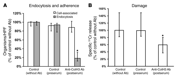
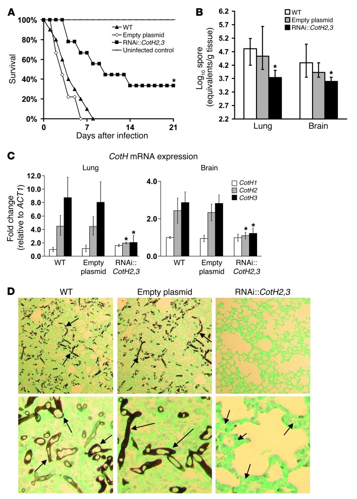
Angioinvasion is a hallmark of mucormycosis. Previously, we identified endothelial cell glucose-regulated protein 78 (GRP78) as a receptor for Mucorales that mediates host cell invasion. Here we determined that spore coat protein homologs (CotH) of Mucorales act as fungal ligands for GRP78. CotH proteins were widely present in Mucorales and absent from noninvasive pathogens. Heterologous expression of CotH3 and CotH2 in Saccharomyces cerevisiae conferred the ability to invade host cells via binding to GRP78. Homology modeling and computational docking studies indicated structurally compatible interactions between GRP78 and both CotH3 and CotH2. A mutant of Rhizopus oryzae, the most common cause of mucormycosis, with reduced CotH expression was impaired for invading and damaging endothelial cells and CHO cells overexpressing GRP78. This strain also exhibited reduced virulence in a diabetic ketoacidotic (DKA) mouse model of mucormycosis. Treatment with anti-CotH Abs abolished the ability of R. oryzae to invade host cells and protected DKA mice from mucormycosis. The presence of CotH in Mucorales explained the specific susceptibility of DKA patients, who have increased GRP78 levels, to mucormycosis. Together, these data indicate that CotH3 and CotH2 function as invasins that interact with host cell GRP78 to mediate pathogenic host-cell interactions and identify CotH as a promising therapeutic target for mucormycosis.


Figures

Figure 1
Figure 1. Identification and expression of CotH genes.
(A) Far-Western blot of R. oryzae surface proteins that bound to GRP78. (B) Dendrogram showing the close identity between CotH2 and CotH3 predicated proteins and the divergence of the CotH proteins from RO3G_16295, the fourth identified ORF widely present in fungi without an identified function. (C) All 3 CotH genes were expressed in resting spores, but only CotH3 was expressed in germlings, of R. oryzae grown in YPD at 37°C. ACT1 represents the loading control. Lanes were run on the same gel but were noncontiguous. (D) Exposure of R. oryzae germlings to endothelial cells induced expression of only CotH2 and CotH3, as determined by RT-PCR. (E) Quantification of CotH mRNA expression in R. oryzae germlings on endothelial cells by qRT-PCR (relative to ACT1) demonstrated 16- and 4-fold increases in CotH3 and CotH2 expression, respectively, whereas CotH1 had no detectable expression. RO3G_16295 was not expressed under any condition tested. **P < 0.001 vs. CotH1 and CotH2, Wilcoxon rank-sum test. n = 9 from 3 independent experiments.
Figure 2
Figure 2. S. cerevisiae cells heterologously expressing CotH2 or CotH3, but not CotH1, interact with host cells through GRP78.
(A) Endothelial cell surface proteins were labeled with NHS-biotin, then extracted with n-octyl-β-d-glucopyranoside in PBS-CM and protease inhibitors. Labeled proteins (250 mg) were incubated with 2 × 10⁸ S. cerevisiae cells, after which unbound proteins were removed by extensive rinsing with PBS-CM. The membrane proteins that remained bound to the organisms were eluted with 6 M urea, separated on 10% SDS-PAGE, and identified by immunoblotting with anti-GRP78 Ab. (B and C) Adherence and endocytosis (determined by differential fluorescence) assays were carried out using endothelial cells (B) or GRP78-overexpressing or parent CHO cells (C) split on 12-mm glass coverslips. HPF, high-power field. *P < 0.001 vs. empty plasmid or CotH1, **P < 0.001 vs. CotH1 and CotH2, Wilcoxon rank-sum test. n = 9 from 3 independent experiments. Data are median ± interquartile range.
Figure 3
Figure 3. GRP78 on intact endothelial cells colocalizes with R. oryzae expressing CotH proteins during endocytosis.
Confocal microscopic images of endothelial cells infected with empty plasmid–transformed (A and C) or CotH2/CotH3 RNAi construct–transformed (B and D) R. oryzae that were germinated for 1 hour. Confluent endothelial cells on a 12-mm-diameter glass coverslip were infected with 105 R. oryzae germlings. After a 40-minute incubation at 37°C, cells were fixed with 3% paraformaldehyde, washed, blocked, and then stained for GRP78 and CotH. Merged images show colocalization (yellow). The same field was taken with differential interference contrast (DIC) imaging to show the presence of R. oryzae. Arrows indicate GRP78 accumulation, CotH cell surface staining, and GRP78/CotH colocalization on R. oryzae. Scale bars: 30 μm (A and B); 15 μm (C and D).
Figure 4
Figure 4. Anti-CotH3 Abs block endothelial cell endocytosis of and damage by R. oryzae.
Endothelial cells were incubated with 50 μg/ml anti-CotH3, with serum from the same rabbit prior to vaccination (preserum control), for 1 hour prior to addition of R. oryzae germlings. (A) Adherence and endocytosis (determined by differential fluorescence) assays were carried out using endothelial cells split on 12-mm glass coverslips. Blocking of CotH3 and CotH2 (since the Abs react to CotH2 proteins) abrogated endocytosis of R. oryzae by endothelial cells. Data were derived from >700 fungal cells interacting with approximately 200 endothelial cells/group/experiment, with an average of 59% cells being endocytosed in the control. (B) Damage was carried out using the 96-well plate 51Cr release method. Blocking of CotH3 and CotH2 reduced the ability of R. oryzae to cause endothelial cell damage. *P < 0.01 vs. either control, Wilcoxon rank-sum test. n = 6 slides or wells per group from 3 (A) or 2 (B) independent experiments. Data are median ± interquartile range.
Figure 5
Figure 5. RNAi targeting CotH2 and CotH3 inhibits the expression of both genes and reduces CotH2 and CotH3 protein synthesis on the cell surface.
R. oryzae was transformed with an RNAi construct targeting CotH2 and CotH3 expression or with empty plasmid. (A) 2 transformants demonstrated >80% reduction in CotH2 and CotH3 expression relative to empty plasmid–transformed R. oryzae, as determined by qRT-PCR. (B) Flow cytometry testing using anti-CotH Abs demonstrated reduced cell surface expression of CotH proteins on RNAi-transformed R. oryzae compared with empty plasmid–transformed, wild-type, or negative control (stained with commercial IgG instead of anti-CotH Abs) R. oryzae. *P < 0.001, #P < 0.005 vs. empty plasmid, Wilcoxon rank-sum test. n = 6 per group from 2 independent experiments.
Figure 6
Figure 6. Inhibition of CotH2 and CotH3 expression decreases the ability of R. oryzae to invade and damage endothelial cells and GRP78-overexpressing CHO cells.
(A and B) RNAi-transformed R. oryzae germlings caused less invasion (A) and damage (B) to endothelial cells compared with empty plasmid–transformed R. oryzae. (C) RNAi-transformed R. oryzae caused equivalent damage to GRP78-overexpressing versus parent CHO cells. Conversely, wild-type or empty plasmid–transformed R. oryzae germlings caused significantly more damage to GRP78-overexpressing cells. *P < 0.005 vs. empty plasmid, **P < 0.01 vs. wild-type and empty plasmid, P < 0.01 vs. parent cells, Wilcoxon rank-sum test. n = 6 slides/group (A) or 9 wells/group (B) from 3 independent experiments. Data are median ± interquartile range.
Figure 7
Figure 7. Comparative binding models of CotH proteins to GRP78.
Models of CotH proteins were generated by homology threading from templates with known structures determined by crystallography. Molecular docking was then evaluated using CotH protein as the ligand and GRP78 as the target. The visualized water-accessible surface areas and secondary structures were energy-minimized for CotH3 (yellow), CotH2 (beige), or CotH1 (white) binding to GRP78 (green). Comparative contact areas of respective CotH proteins are shown in blue. Note the large, bimodal binding cleft in CotH3 that was also partially present in CotH2, consistent with biological findings. In contrast, CotH1 exhibited a limited contact area on a distinct aspect of the GRP78 target protein. Of interest are the comparative structural domains that may confer GRP78 interactions by differing CotH proteins. For example, each of the CotH proteins has multiple α-helical domains that appear to serve as a structural scaffold. However, the proteins differ considerably in the extent of their β-sheet conformation. For example, CotH3 exhibits an extended docking-predicted GRP78 contact facet, with little β-sheet stabilization. Conversely, CotH2 and, to a lesser extent, CotH1 contain explicit β-sheet structural domains. In the former case, this domain forms a component of the GRP78-docking facet. In the latter, the β-sheet domain does not appear to be involved in interactions with GRP78. Thus, despite similarities in their overall topology, more specific 3D aspects likely determine the capability for distinct CotH protein interactions with GRP78.
Figure 8
Figure 8. Inhibition of CotH2 and CotH3 expression attenuates R. oryzae virulence in DKA mice.
(A) Survival of mice infected intratracheally with wild-type (n = 10), empty plasmid–transformed (n = 9), and RNAi-transformed (n = 9) R. oryzae (inhaled inocula, 2.4 × 10³, 2.8 × 10³, and 2.5 × 10³ spores, respectively). *P < 0.003 vs. wild-type and empty plasmid, log-rank test. (B) Lungs and brain fungal burden of DKA mice (n = 9 per group) infected intratracheally with wild-type, empty plasmid–transformed, or RNAi-transformed cells (1.7 × 10³, 3.0 × 10³, and 3.1 × 10³, respectively). Mice were sacrificed on day +2 relative to infection, and their organs were processed for tissue fungal burden, determined by qPCR. Data are median ± interquartile range. *P < 0.001 versus wild-type and empty plasmid, Wilcoxon rank-sum test. (C) In vivo expression of CotH genes in lungs and brains harvested from mice infected with wild-type, empty plasmid–transformed, or RNAi-transformed R. oryzae, as determined by qRT-PCR using specific primers to each CotH gene. Data are mean ± SD. *P < 0.001 vs. wild-type and empty plasmid. (D) PAS-stained sections of lungs demonstrated extensive hyphal elements (arrows) and signs of severe pneumonia from organs collected from DKA mice infected with wild-type or empty plasmid–transformed R. oryzae, whereas mild pneumonia (arrows) was observed in lungs from mice infected with RNAi-transformed R. oryzae. Original magnification, ×100 (top); ×400 (bottom).
Figure 9
Figure 9. Anti-CotH Abs protect DKA mice from R. oryzae infection.
DKA mice were treated with 1 mg purified IgG collected from rabbit serum after vaccination with CotH3 peptide. Abs were administered intraperitoneally to mice either 2 hours before (A) or 24 hours after (B) infecting them intratracheally with 2.5 × 10⁵ wild-type R. oryzae spores. Average delivered inoculum to the lungs was 6.0 × 10³ (A) and 3.8 × 10³ (B) spores. n = 14 (A), 10 (B) per arm. *P = 0.005, **P = 0.02, log-rank test.

Comment in

Similar articles

Cited by

References

    1. Sugar A. Principles and Practice of Infectious Diseases. Agents of mucormycosis and related species. In: Mandell GL, Bennett JE, Dolin R, eds. Philadelphia, Pennsylvania, USA: Elsevier; 2005:2979.
    1. Ibrahim AS, Edwards JJE, Filler SG, Spellberg B. Essentials of Clinical Mycology. 2011. Mucormycosis and entomophthoramycosis (zygomycosis). pp. 265–280. In: Kauffman CA, Pappas PG, Sobel JD, Dismukes WE, eds. New York, New York, USA: Springer;
    1. Ribes J, Vanover-Sams CL, Baker DJ. Zygomycetes in human disease. Clin Microbiol Rev. 2000;13(2):236–301. doi: 10.1128/CMR.13.2.236-301.2000. - DOI - PMC - PubMed
    1. Ibrahim A. Host cell invasion in mucormycosis: role of iron. Curr Opin Microbiol. 2011;14(4):406–411. doi: 10.1016/j.mib.2011.07.004. - DOI - PMC - PubMed
    1. Spellberg B, Walsh TJ, Kontoyiannis DP, Edwards J, Jr, Ibrahim AS. Recent advances in the management of mucormycosis: from bench to bedside. Clin Infect Dis. 2009;48(12):1743–1751. doi: 10.1086/599105. - DOI - PMC - PubMed

Publication types

MeSH terms