Review
. 2014 Jan;15(1):34-48.
doi: 10.1038/nrm3723.

Cell intercalation from top to bottom

Affiliations
Review

Cell intercalation from top to bottom

Elise Walck-Shannon et al. Nat Rev Mol Cell Biol. 2014 Jan.

Abstract

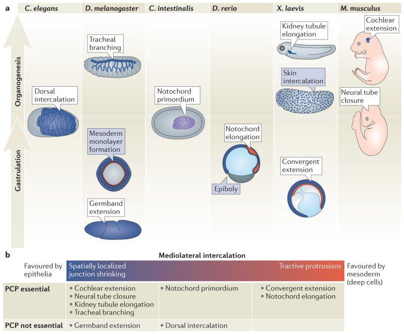
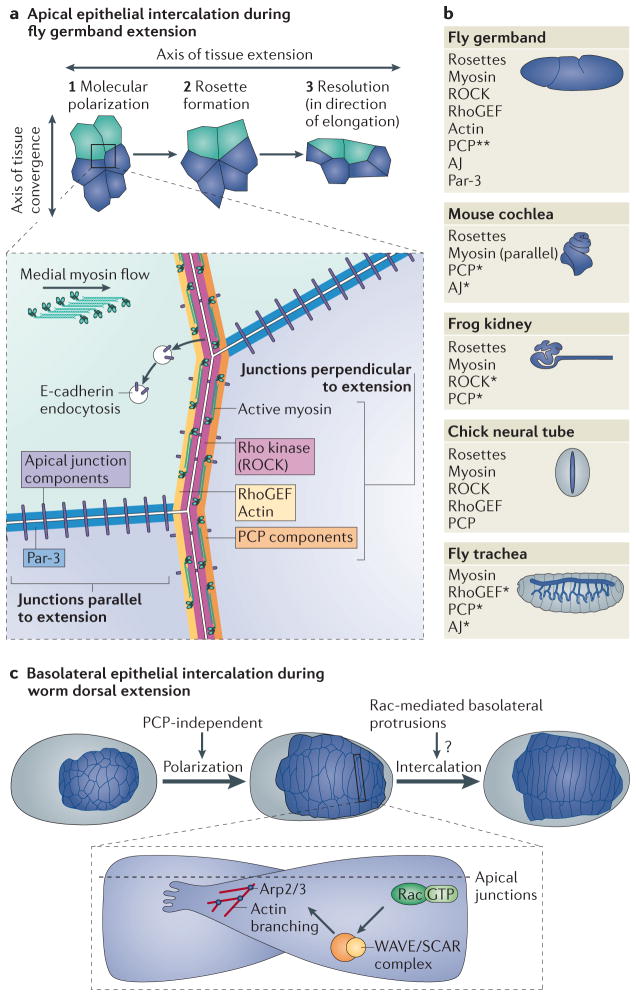
Animal development requires a carefully orchestrated cascade of cell fate specification events and cellular movements. A surprisingly small number of choreographed cellular behaviours are used repeatedly to shape the animal body plan. Among these, cell intercalation lengthens or spreads a tissue at the expense of narrowing along an orthogonal axis. Key steps in the polarization of both mediolaterally and radially intercalating cells have now been clarified. In these different contexts, intercalation seems to require a distinct combination of mechanisms, including adhesive changes that allow cells to rearrange, cytoskeletal events through which cells exert the forces needed for cell neighbour exchange, and in some cases the regulation of these processes through planar cell polarity.


Conflict of interest statement

Competing interests statement: The authors declare no competing interests.

Figures

Figure 1
Figure 1. Intercalation events drive morphogenesis in diverse contexts during metazoan development
a, Intercalation events promote morphogenesis during distinct stages of development, including gastrulation and organogenesis (shown vertically in the figure). These include mediolateral intercalation events (in white boxes) and radial intercalation events (blue boxes). In all figures, epithelial structures are in blue; mesodermal structures are in red; endoderm structures are not shown. The C. intestinalis (ascidian) notochord is unusual in that it behaves as an epithelium early and later like a non-epithelial mesoderm, and thus is shown in purple. Embryos are shown laterally with anterior to the left and posterior to the right, except C. elegans and C. intestinalis which are shown as dorsal views, the Drosophila mesoderm, which is shown as a cross-sectional view, and the mouse embryo, which is shown as anterior up and posterior down. b, Mediolateral intercalation can be driven by different cellular programmes. Many epithelia undergo cell intercalation through tight regulation of their apical junctions, whereas many mesodermal cells undergo intercalation through basolateral protrusive activity. However, these mechanisms may represent two poles along a continuum, with some intercalation events displaying only some characteristics of each pole. The different intercalation events are listed below, at a position that reflects the mechanisms that drive mediolateral intercalation in this context. Only a subset of these require planar cell polarity (PCP) signalling.
Figure 2
Figure 2. Intercalation in epithelial cells can be driven through junction remodelling or protrusion formation
a, During germband extension in D. melanogaster, epithelial mediolateral intercalation relies on shrinkage of junctions that lie perpendicular to the axis of extension. Junction disassembly often results in rosette formation, which resolves in the direction of tissue extension. Cartoon cells were traced from live images of GBE cells, available at http://www.hhmi.org/research/molecular-control-polarized-cell-behavior. Junctions parallel to the direction of extension are enriched for apical junction components and Par-3 (inset). By contrast, junctions perpendicular to the direction of tissue extension are enriched for planar cell polarity (PCP) components, and shrinking of these junctions is driven by myosin, and—in some cases—cadherin. Non-muscle myosin II, F-actin, ROCK and RhoGEF2 all accumulate specifically at shrinking vertical junctions. E-cadherin endocytosis occurs at perpendicular junctions, which may contribute to reduced adhesion there. Medial myosin flow also correlates with junction shrinkage. Comparison between intercalation events during fly germband extension and in epithelial tubes, including the mouse cochlea, frog kidney, chick neural tube, and fly trachea. An asterisk (*) indicates that a requirement for that component has been demonstrated, but its junctional polarization has not yet been determined and a double asterisk (**) indicates that the component has a polarized distribution, but is not required for intercalation. The remaining factors shown seem to be both required and asymmetrically polarized. All the systems shown, except the fly trachea, require rosette formation for intercalation. c, Basolateral protrusion formation during dorsal intercalation in C. elegans. Cells extend tips towards the midline and basal to the apical junctions, through Rac-dependent regulation of actin branching via the nucleating complex Arp2/3. Intercalation is completed once the tips reach the lateral (seam) cell on the contralateral side. Polarization of these cells appears to be PCP-independent.
Figure 3
Figure 3. Mediolateral intercalation of deep mesodermal cells in vertebrates
a, Ascidian notochord intercalation requires both planar cell polarity (PCP) and the basement membrane. Cells in the notochord primordium, which is initially epithelial (purple), extend basolateral protrusions (BLPs) along the apico(A)-basal(B) axis (left, inset). The notochord eventually behaves more like mesoderm and is polarized by the PCP pathway, which works in parallel to the basement membrane to direct completion of intercalation. b, Mesodermal mediolateral intercalation requires downregulation of C-cadherin. In the example shown here, during chordamesoderm convergent extension in a frog Keller explant, cells extend bidirectional, mediolateral protrusions, which provide traction for cell intercalation towards the midline. Inset: At the mediolateral ends of intercalating cells, PAPC and Frizzled bound to C-cadherin inhibit cell adhesion by inhibiting C-cadherin clustering, while both PAPC and the PCP pathway help to activate cytoskeletal extensions through regulation of the RhoGTPases Rac and Rho and Arp2/3-mediated actin nucleation. PAPC may activate Rho by inhibiting the transcription of Rho GAPs. PAPC that is not bound to Frizzled undergoes endocytosis. The extracellular matrix (ECM) component fibronectin (FN) also signals to more apical pathways through interactions with integrin and synedcan. c, Later phases of mediolateral intercalation in the notochord. Boundary formation requires laminin β1 and γ1 in zebrafish (yellow) and completion of intercalation requires the PCP pathway. The PCP components Prickle and Dishevelled (Dsh) localize to puncta at the anterior (A) and posterior (P) sides of the cell, respectively (inset, right).
Figure 4
Figure 4. Radial intercalation drives morphogenesis during gastrulation and later in development
a, Radial intercalation of the Drosophila mesoderm (red) requires long-range FGF signals from the ectoderm to polarize radial protrusions. These radial protrusions extend into the mesoderm, where they promote E-cadherin-mediated adhesions between mesodermal cells. There is still some question about the involvement of integrin, which is expressed by mesodermal cells, in this process. b, Zebrafish epiboly depends on radial intercalation of deep cells (green) underneath the enveloping layer (EVL). Cell adhesion mediated by EpCAM in the EVL and E-cadherin in the deep cells and/or the EVL are required for intercalation. E-cadherin is regulated by the G protein Gα12/13. Strong E-cadherin attachment to the cytoskeleton, via α-catenin inhibits blebbing, allowing for efficient radial intercalation. The EGF pathway, which is downstream of the transcription factor Pou5f1, is required for proper E-cadherin endocytosis and normal intercalation. E-cadherin-mediated adhesion counterbalances α-catenin/Ezrin-mediated blebbing in deep cells. c, Radial intercalation in the Xenopus prechordal plate mesoderm also relies on long range signals. PDGF-A, secreted from the overlying blastocoel roof is required for radial orientation of cell protrusions and radial intercalation. Intercalation fails when PDGF-A is depleted by morpholinos (MO). d, Radial intercalation of ciliated cell precursors (CCPs) into the outer superficial layer in Xenopus skin depends both cell-autonomously on factors to specify the apical edge (including Rab11 recycling) and cell-non-autonomously on factors that bind to the extracellular matrix (including dystroglycan, which increases levels of E-cadherin, fibronectin and laminin). After CCP radial intercalation, it matures into a ciliated cell.

Similar articles

Cited by

References

Works Cited

    1. Keller R. Developmental biology. Physical biology returns to morphogenesis. Science. 2012;338:201–3. - PubMed
    1. Keller R. Mechanisms of elongation in embryogenesis. Development. 2006;133:2291–2302. - PubMed
    1. Wallingford JB. Planar cell polarity and the developmental control of cell behavior in vertebrate embryos. Annu Rev Cell Dev Biol. 2012;28:627–53. - PubMed
    1. Carroll TJ, Yu J. The kidney and planar cell polarity. Curr Top Dev Biol. 2012;101:185–212. - PMC - PubMed
    1. Tada M, Kai M. Planar cell polarity in coordinated and directed movements. Curr Top Dev Biol. 2012;101:77–110. - PubMed

Highlighted References

    1. Levayer R, Lecuit T. Oscillation and Polarity of E-Cadherin Asymmetries Control Actomyosin Flow Patterns during Morphogenesis. Developmental Cell. 2013 doi: 10.1016/j.devcel.2013.06.020. - DOI - PubMed
    1. Fernandez-Gonzalez R, Simoes S, de M, Röper J-C, Eaton S, Zallen JA. Myosin II dynamics are regulated by tension in intercalating cells. Developmental cell. 2009;17:736–43. - PMC - PubMed
    1. Nishimura T, Honda H, Takeichi M. Planar cell polarity links axes of spatial dynamics in neural-tube closure. Cell. 2012;149:1084–97. - PubMed
    1. Lienkamp SS, et al. Vertebrate kidney tubules elongate using a planar cell polarity-dependent, rosette-based mechanism of convergent extension. Nature Genetics. 2012;44:1382–7. - PMC - PubMed
    1. Chacon-Heszele MF, Ren D, Reynolds AB, Chi F, Chen P. Regulation of cochlear convergent extension by the vertebrate planar cell polarity pathway is dependent on p120-catenin. Development. 2012;139:968–78. - PMC - PubMed

Publication types

LinkOut - more resources