

Review
doi: 10.1038/nrn3650.
Branch management: mechanisms of axon branching in the developing vertebrate CNS
Affiliations
- PMID: 24356070
- PMCID: PMC4063290
- DOI: 10.1038/nrn3650
Item in Clipboard
Review
Branch management: mechanisms of axon branching in the developing vertebrate CNS
Nat Rev Neurosci.
2014 Jan.
Abstract
The remarkable ability of a single axon to extend multiple branches and form terminal arbors enables vertebrate neurons to integrate information from divergent regions of the nervous system. Axons select appropriate pathways during development, but it is the branches that extend interstitially from the axon shaft and arborize at specific targets that are responsible for virtually all of the synaptic connectivity in the vertebrate CNS. How do axons form branches at specific target regions? Recent studies have identified molecular cues that activate intracellular signalling pathways in axons and mediate dynamic reorganization of the cytoskeleton to promote the formation of axon branches.
Figures
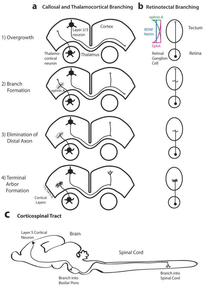
In the mammalian corpus callosum and thalamocortical pathway (a) and in the chick retinotectal pathway (b), axons initially extend past their eventual terminal regions (1). After a delay, branches extend interstitially from the axon shaft (2) followed by elimination of the distal axon (3) and formation of terminal arbors in topographically correct target regions (4). Figure c depicts interstitial branching from an individual layer 5 sensorimotor cortical axon into the pons and spinal cord (adapted from ). In the retinotectal system (b), opposing gradients of EPHA–ephrin repellents inhibit retinal axon branching anterior and posterior to the correct target, whereas positive BDNF cues along the tectum promote branching at appropriate topographic positions, . Callosal axons connect homotypic regions of the cortical hemispheres but can also branch to other cortical areas, (not shown). Thalamocortical axons form branches that arborize topographically in layer 4 of somatosensory cortex but other thalamocortical axons can branch into multiple cortical areas (not shown).
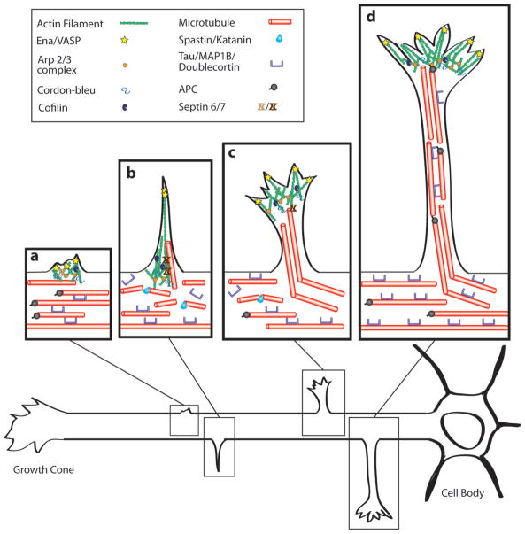
a | Membrane protrusion requires the accumulation of actin filaments, which can form patches–. In (a) membrane protrusion requires actin nucleation (ARP2/3, and Cordon-bleu), actin branching (ARP2/3) and actin elongation (Ena/VASP). Cofilin is important for the turnover of actin filaments and Septin 6 is localized to the actin patch. MTs are stabilized by MAPs (Tau//Doublecortin),, in the axon but are also capable of extending at their plus ends (APC/EB proteins),. b | Filopodia, which contain bundled actin filaments, emerge from the axon shaft accompanied by localized splaying and fragmentation of MTs,,,,; these invade the filopodium and extend along actin bundles. MTs are severed by Spastin and Katanin. Septin 7 promotes the entry of MTs into the filopodium. c, d | Stable MTs enter the nascent branch (c), which continues to mature and extend; this growth is led by a motile growth cone (c, d). MAPs protect some of the MTs in the axon shaft from severing (c, d). Within the axon branch MTs become bundled and stabilized by MAPs (d).
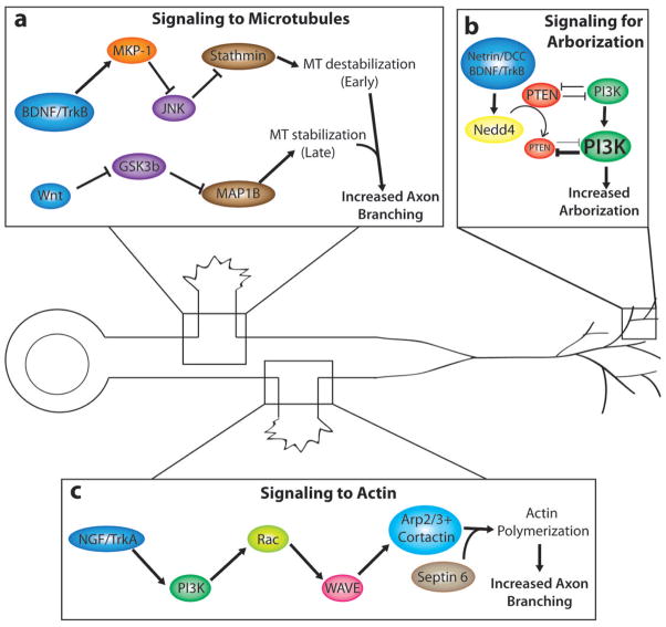
a | Different pathways leading to MT destabilization (top) at early branching stages or by MT stabilization at later branching stage (bottom) can each promote axon branching by opposing effects on MT stability. Differential effects on MT stability are shown in two examples of branching induced by BDNF and its TRKB receptor (top) in cortical neurons or the morphogen WNT7A (bottom) in pontine mossy fibers,
b | BDNF and netrins increase terminal arborization of frog retinotectal axons, . In this signalling pathway, NEDD4 ubiquitinates PTEN and PTEN degradation leads to increased terminal axon branching by promotion of PI3K, which is known to regulate cytoskeletal dynamics. By contrast, inhibition of NEDD4 leads to an increase in PTEN levels and hence inhibition of axon branching. c | NGF-induced TRKA signalling promotes axon branching in chick sensory (DRG) axons. It does this by activating PI3K and, in turn, RAC, which activates actin associated proteins to increase actin polymerization and the formation of actin patches. Cortactin, recruited by Septin6, promotes the emergence of filopodia from actin patches. Abbreviations: brain-derived neurotrophic factor (BDNF), mitogen activated protein kinase phosphatase (MKP-1), MAPK c-jun N-terminal kinase (JNK), stathmin (STMN), phosphoinositide 3-kinase (PI3K), ubiquitin ligase E3 (NEDD4), the phosphatase phosphatidylinositol 3,4,5 trisphosphate (PTEN), nerve growth factor (NGF), actin related protein (ARP 2/3).
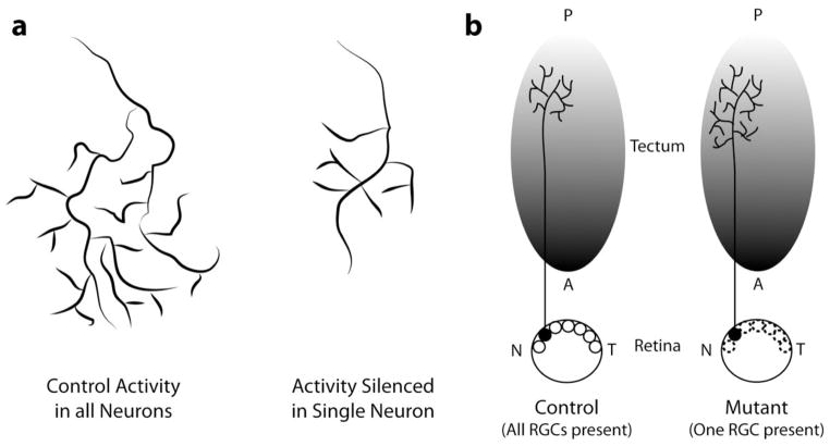
a | The terminal arbor of a retinal axon in the zebrafish optic tectum with normal electrical activity has a complex highly branched morphology (left), and it can outcompete a neighbouring retinotectal arbor in which electrical activity has been silenced (right), resulting in a smaller less elaborate morphology in the silenced axon. b | A retinal axon arbor in a normal control zebrafish tectum (left) has a smaller less complex morphology when all other retinal gangion cells (RCGs) are present. In the mutant zebrafish with only a single RCG axon (right), arbors terminate in appropriate tectal regions but are larger and more complex in the absence of competitive interactions with neighboring axons (figure adapted from ). Anterior posterior tectum (A, P), nasal temporal retina (N, T).
References
-
- Acebes A, Ferrus A. Cellular and molecular features of axon collaterals and dendrites. Trends Neurosci. 2000;23:557–65. - PubMed
-
- Gallo G. The cytoskeletal and signaling mechanisms of axon collateral branching. Dev Neurobiol. 2011;71:201–20. - PubMed
-
- Kornack DR, Giger RJ. Probing microtubule +TIPs: regulation of axon branching. Curr Opin Neurobiol. 2005;15:58–66. - PubMed
Publication types
MeSH terms
Grants and funding
LinkOut - more resources
Full Text Sources
Other Literature Sources
