. 2014 Mar-Apr;20(2):129-35.
doi: 10.5152/dir.2013.13236.

Follow-up of true visceral artery aneurysm after coil embolization by three-dimensional contrast-enhanced MR angiography

Affiliations

Follow-up of true visceral artery aneurysm after coil embolization by three-dimensional contrast-enhanced MR angiography

Masamichi Koganemaru et al. Diagn Interv Radiol. 2014 Mar-Apr.

Abstract

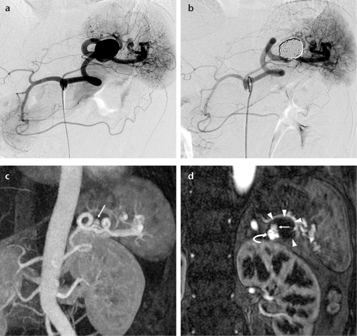
Purpose: We aimed to evaluate the outcomes of coil embolization of true visceral artery aneurysms by three-dimensional contrast-enhanced magnetic resonance (MR) angiography.

Materials and methods: We used three-dimensional contrast-enhanced MR angiography, which included source images, to evaluate 23 patients (mean age, 60 years; range, 28-83 years) with true visceral artery aneurysms (splenic, n=15; hepatic, n=2; gastroduodenal, n=2; celiac, n=2; pancreaticoduodenal, n=1; gastroepiploic, n=1) who underwent coil embolization. Angiographic aneurysmal occlusion was revealed in all cases. Follow-up MR angiography was conducted with either a 1.5 or 3 Tesla system 3-25 months (mean, 18 months) after embolization. MR angiography was evaluated for aneurysmal occlusion, hemodynamic status, and complications.

Results: Complete aneurysmal occlusion was determined in 22 patients (96%) on follow-up MR angiography (mean follow-up period, 18 months). Neck recanalization, which was observed at nine and 20 months after embolization, was confirmed in one of eight patients (13%) using a neck preservation technique. In this patient, a small neck recanalization covered by a coil mass was demonstrated. The complete hemodynamic status after embolization was determined in 21 patients (91%); the visualization of several collateral vessels, such as short gastric arteries, after parent artery occlusion was poor compared with that seen on digital subtraction angiography in the remaining two patients (9%). An asymptomatic localized splenic infarction was confirmed in one patient (4%).

Conclusion: Our study presents the follow-up results from three-dimensional contrast-enhanced MR angiography, which confirmed neck recanalization, the approximate hemodynamic status, and complications. This effective and less invasive method may be suitable for serial follow-up after coil embolization of true visceral aneurysms.


Figures

Figure 1. a–d.
Figure 1. a–d.
A 47-year-old female with a true splenic artery aneurysm. A celiac arteriogram (a) shows a saccular splenic artery aneurysm. A celiac arteriogram after coil embolization using a neck preservation technique (b) shows occlusion of the aneurysm and patency of the splenic artery. Follow-up contrast-enhanced 1.5 T MR angiograms (maximum intensity projection and source image) performed at nine months after initial embolization (c, d) show clear enhancement of the enlarged parent artery (d, curved arrow) without an increase in aneurysm size (d, arrowheads). This suggested neck recanalization (c, d, arrows).
Figure 2. a–e.
Figure 2. a–e.
A 69-year-old male with a true celiac artery aneurysm. A celiac arteriogram after coil embolization using the coil packing technique (a) shows complete occlusion of the aneurysm (curved arrow) and the common hepatic artery via collateral circulation by the right gastric artery from the left gastric artery (arrows). A superior mesenteric arteriogram after coil embolization (b) shows complete occlusion of the aneurysm (curved arrow). Note the arterial flow of the hepatic arteries via collateral circulation by the pancreaticoduodenal arteries (arrowheads) and the splenic artery via collateral circulation by the dorsal pancreatic artery (arrow). Unenhanced CT for detailed examination of acute cholangitis four months after coil embolization (c) shows strong metallic coil artifacts. Follow-up contrast-enhanced 3 T MRI (dynamic study; axial image) performed three months after initial embolization (d) shows complete occlusion of the aneurysm without metallic artifacts (arrowheads). Follow-up contrast-enhanced 3 T MR angiogram (maximum intensity projection; oblique view) performed three months after initial embolization (e) shows clear enhancement of the collateral circulation, such as the right gastric artery (white arrows), pancreaticoduodenal arteries (arrowheads), and dorsal pancreatic artery (black arrow).
Figure 3. a, b.
Figure 3. a, b.
A 57-year-old male with a true splenic artery aneurysm. A celiac arteriogram after coil embolization using coil packing and the isolation technique (a) shows complete occlusion of the aneurysm (curved arrows). Note the arterial reperfusion of the spleen via collateral circulation by the short gastric arteries from the left gastric artery (a, arrowheads) and the gastroepiploic artery from the gastroduodenal artery (a, arrows). A follow-up contrast-enhanced MR angiogram (maximum intensity projection) using 3 T MRI performed 22 months after initial embolization (b) shows complete occlusion of the aneurysm and arterial reperfusion of the spleen via collateral circulation by the gastroepiploic artery (arrows). Other collateral vessels, such as short gastric arteries, are not shown.
Figure 4. a–d.
Figure 4. a–d.
A 69-year-old male with a true inferior pancreaticoduodenal artery aneurysm. A superior mesenteric arteriogram (a) shows a saccular inferior pancreaticoduodenal artery aneurysm (arrow). A superior mesenteric arteriogram after coil embolization using the coil packing technique (b) shows occlusion of the aneurysm (arrow). Follow-up contrast-enhanced 3 T MRI (dynamic study; axial image) performed 22 months after initial embolization (c) shows complete occlusion of the aneurysm without metallic artifacts (arrowheads). A follow-up contrast-enhanced MR angiogram (maximum intensity projection) using 3 T MRI performed 22 months after initial embolization shows (d) complete occlusion of the aneurysm (black arrow) and clear enhancement of the pancreaticoduodenal and hepatic arteries (white arrows).

References

    1. Carmeci C, McClenathan J. Visceral artery aneurysms as seen in a community hospital. Am J Surg. 2000;179:486–489. - PubMed
    1. Kalko Y, Ugurlucan M, Basaran M, et al. Visceral artery aneurysms. Heart Surg Forum. 2007;10:E24–29. - PubMed
    1. Henke PK, Cardneau JD, Welling TH, 3rd, et al. Renal artery aneurysms: a 35-year clinical experience with 252 aneurysms in 168 patients. Ann Surg. 2001;234:454–462. - PMC - PubMed
    1. Shanley CJ, Shah NL, Messina LM. Common splanchnic artery aneurysms: splenic, hepatic, and celiac. Ann Vasc Surg. 1996;10:315–322. - PubMed
    1. Wagner WH, Allins AD, Treiman RL, et al. Ruptured visceral artery aneurysms. Ann Vasc Surg. 1997;11:342–347. - PubMed

Publication types