. 2014 Feb;25(4):495-507.
doi: 10.1091/mbc.E13-09-0526. Epub 2013 Dec 19.

The novel centriolar satellite protein SSX2IP targets Cep290 to the ciliary transition zone

Affiliations

The novel centriolar satellite protein SSX2IP targets Cep290 to the ciliary transition zone

Maren Klinger et al. Mol Biol Cell. 2014 Feb.

Abstract

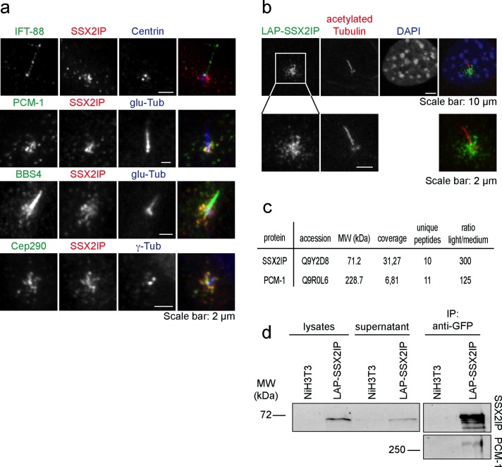
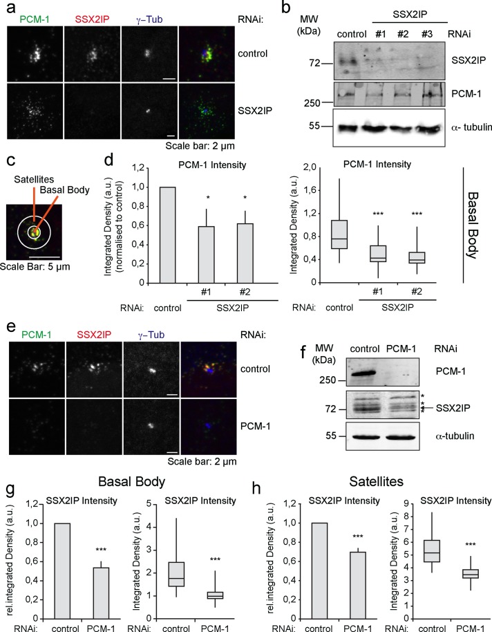
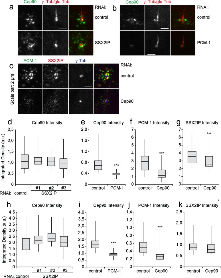
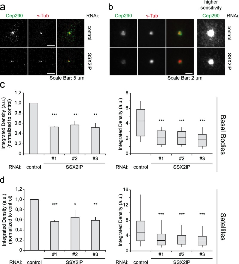
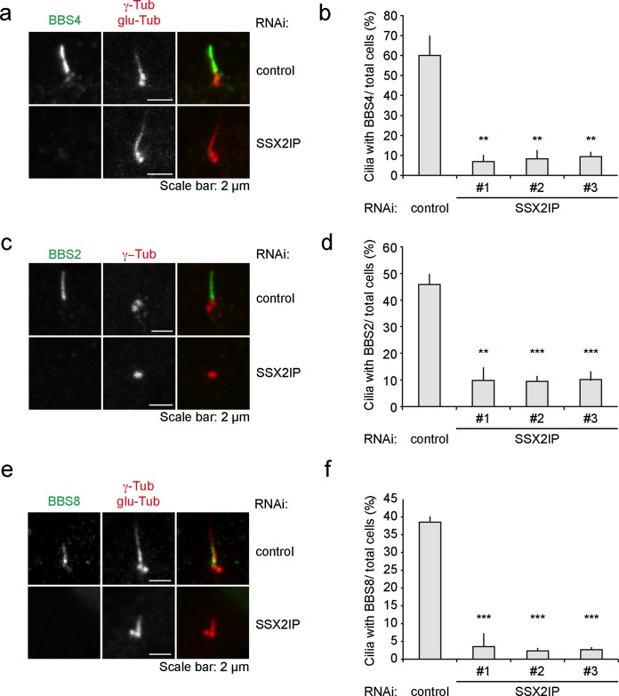
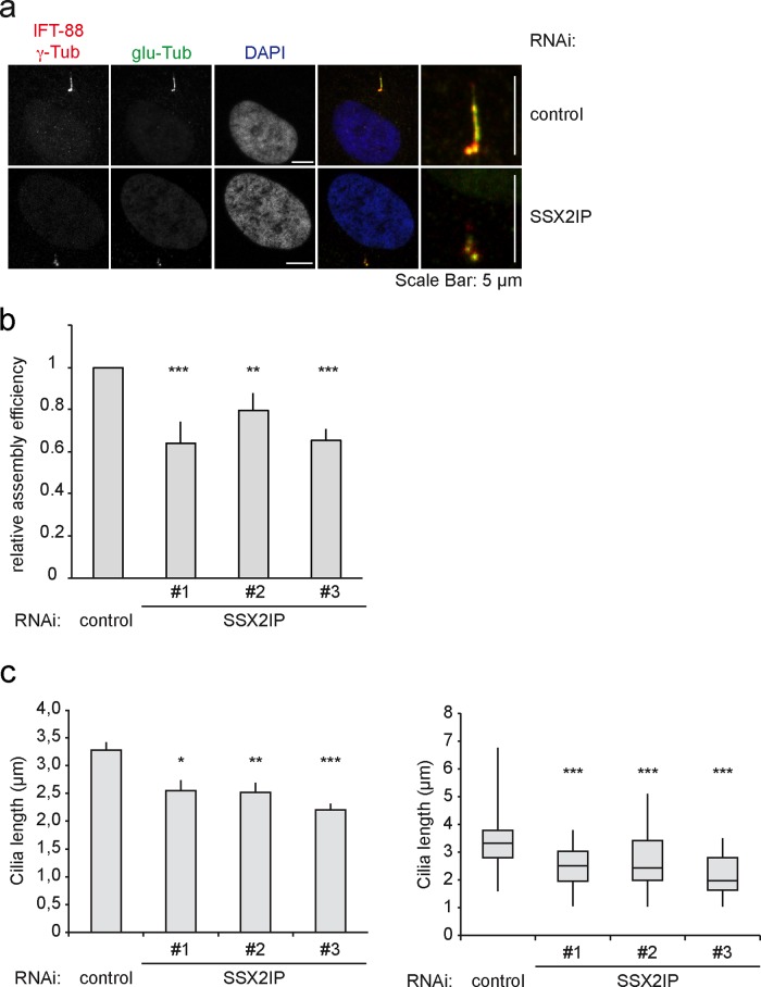
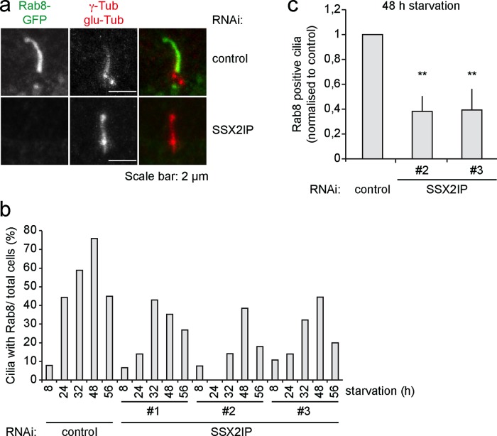
In differentiated human cells, primary cilia fulfill essential functions in converting mechanical or chemical stimuli into intracellular signals. Formation and maintenance of cilia require multiple functions associated with the centriole-derived basal body, from which axonemal microtubules grow and which assembles a gate to maintain the specific ciliary proteome. Here we characterize the function of a novel centriolar satellite protein, synovial sarcoma X breakpoint-interacting protein 2 (SSX2IP), in the assembly of primary cilia. We show that SSX2IP localizes to the basal body of primary cilia in human and murine ciliated cells. Using small interfering RNA knockdown in human cells, we demonstrate the importance of SSX2IP for efficient recruitment of the ciliopathy-associated satellite protein Cep290 to both satellites and the basal body. Cep290 takes a central role in gating proteins to the ciliary compartment. Consistent with that, loss of SSX2IP drastically reduces entry of the BBSome, which functions to target membrane proteins to primary cilia, and interferes with efficient accumulation of the key regulator of ciliary membrane protein targeting, Rab8. Finally, we show that SSX2IP knockdown limits targeting of the ciliary membrane protein and BBSome cargo, somatostatin receptor 3, and significantly reduces axoneme length. Our data establish SSX2IP as a novel targeting factor for ciliary membrane proteins cooperating with Cep290, the BBSome, and Rab8.


Figures

FIGURE 1:
FIGURE 1:
SSX2IP is a centriolar satellite protein localizing to basal bodies in ciliated cells. (a) Indirect immunofluorescence of serum-starved RPE-1 cells with specific antibodies as indicated; glu-tub, glutamylated tubulin. (b) Indirect immunofluorescence in NIH3T3 cells stably expressing LAP-SSX2IP (note that the LAP tag contains GFP), which was visualized with GFP antibodies. (c, d) Immunoprecipitation with single-chain GFP antibodies of lysates from serum-starved NIH3T3 cells and serum-starved NIH3T3 cells stably expressing LAP-SSX2IP. (c) Quantitative mass spectrometry analysis of immunoprecipitates. The N-termini of tryptic peptides were dimethylated using stable carbon isotopes for relative quantification of peptide abundance in SSX2IP immunoprecipitates (light) vs. control samples (medium). (d) Immunoblot shows coimmunoprecipitation of SSX2IP and PCM-1.
FIGURE 2:
FIGURE 2:
SSX2IP stability depends on PCM-1 in RPE-1 cells. Cells were transfected with control, SSX2IP, or PCM-1 siRNA. (a) Indirect immunofluorescence detects PCM-1, SSX2IP, or γ-tubulin after control or SSX2IP siRNA treatment. (b) Immunoblot to document down-regulation of SSX2IP and PCM-1 after down-regulation of SSX2IP; α-tubulin served as a loading control. (c) Schematic view to show the inner (basal body) area (3 μm2) and the outer (satellite) area (19-μm2 ring) quantified using SSX2IP (red) and PCM-1 (green) signals (d, g, h); see Materials and Methods for details. (d) Quantification of signal intensity of PCM-1 at the basal body after control or SSX2IP siRNA treatment. (e) Cells were transfected with control or PCM-1 siRNA and stained for PCM-1, SSX2IP, or γ-tubulin by indirect immunofluorescence. (f) Immunoblot to document siRNA-mediated down-regulation of PCM-1 and down-regulation of SSX2IP after PCM-1 knockdown; α-tubulin served as loading control. The asterisks indicate a cross-reacting band, and the specific signal is marked by the arrow. (g, h) Quantification of signal intensities of SSX2IP at the basal body (g) and in satellites (h) after treatment with control or PCM-1 siRNA. (d, g, h) Left (bars), mean values of average intensities normalized to controls (±SEM) of three independent experiments (n ≥ 150). Right (box-and-whiskers plots), signal distribution of a single representative experiment. *p < 0.05, **p < 0.01, ***p < 0.001 (two-tailed Student's t test).
FIGURE 3:
FIGURE 3:
PCM-1 but not SSX2IP localization is dependent on Cep90 in ciliated RPE-1 cells. (a) Cells were transfected with control or SSX2IP siRNA and stained for Cep90 or γ- tubulin and glutamylated (glu) tubulin using indirect immunofluorescence. (b) Cells were transfected with control or PCM-1 siRNA and stained for of Cep90 or γ-tubulin and glutamylated (glu) tubulin using indirect immunofluorescence. (c) Cells were transfected with control or Cep90s siRNA and stained for PCM-1, SSX2IP, or γ-tubulin using indirect immunofluorescence. (d–g) Quantification of signal intensities at basal bodies. (h–k) Quantification of signal intensities in satellites. (d, h) Cep90 signal intensity after SSX2IP knockdown. (e, i) Cep90 signal intensity after PCM-1 knockdown. (f, j) PCM-1 signal intensity after Cep90 knockdown. (g, k) SSX2IP signal intensity after Cep90 knockdown. n ≥ 150 from one (Cep90) or three experiments. *p < 0.05, **p < 0.01, ***p < 0.001 (two-tailed Student's t test).
FIGURE 4:
FIGURE 4:
Cep290 recruitment to centrosomal satellites and basal bodies depends on SSX2IP in RPE-1 cells. (a) Cells were transfected with control or SSX2IP siRNA and stained for Cep290 and γ-tubulin using indirect immunofluorescence. (b) Average projection of 50 cells immunostained as shown in a. Centering of single images before projection was based on the γ-tubulin signal. (c, d) Quantification of signal intensities of Cep290 at the basal body (c) or in satellites (d) of cells transfected with control or SSX2IP siRNA. (c, d) Left (bars), mean values of averages ± SEM from three independent experiments (n = 150) normalized to controls. Right (box-and-whiskers plots), quantification of a single representative experiment. *p < 0.05, **p < 0.01, ***p < 0.001 (two-tailed Student's t test).
FIGURE 5:
FIGURE 5:
SSX2IP is required for ciliary BBSome localization in RPE-1 cells. (a) Cells were transfected with control or SSX2IPs siRNA and stained for BBS4 or γ-tubulin and glutamylated (glu) tubulin using indirect immunofluorescence. (b) Quantification of immunofluorescence shown in a. Cells with cilia showing ciliary localization of BBS4 were determined in relation to the overall cell number. (c) Cells were transfected with control or SSX2IP siRNA and stained for BBS2 or γ-tubulin and glutamylated (glu) tubulin using indirect immunofluorescence. (d) Quantification of immunofluorescence shown in c. Cells with cilia showing ciliary localization of BBS2 were counted in relation to the overall cell number. (e) Cells were transfected with control or SSX2IP siRNA and stained for BBS2 or γ-tubulin and glutamylated (glu) tubulin using indirect immunofluorescence. (f) Quantification of immunofluorescence shown in c. Cells with cilia showing ciliary localization of BBS2 were counted in relation to the overall cell number. (b, d, f) Mean values from averages from three independent experiments ± SEM (n ≥ 150) normalized to controls. *p < 0.05, **p < 0.01, ***p < 0.001 (two-tailed Student's t test).
FIGURE 6:
FIGURE 6:
SSX2IP knockdown leads to less efficient ciliogenesis and shorter cilia. (a) RPE-1 cells were transfected with control or SSX2IP siRNA and stained for IFT-88, γ-tubulin, or glutamylated (glu) tubulin using indirect immunofluorescence. Magnified merges do not display DAPI staining. (b) Quantification of the relative cilia assembly activity in control and SSX2IP-knockdown cells. Bars show mean values ± SEM from three independent experiments (n ≥ 100 cells). (c) Quantification of cilia lengths in RPE-1 cells after SSX2IP knockdown using the IFT-88, γ-tubulin, and glutamylated tubulin signals. Left (bars), mean values of averages ± SEM from three independent experiments (n ≥ 150) normalized to controls. Right (box-and-whiskers plots), quantification of a single representative experiment. *p < 0.05, **p < 0.01, ***p < 0.001 (two-tailed Student's t test).
FIGURE 7:
FIGURE 7:
Ciliary accumulation of Rab8 is compromised after SSX2IP knockdown in RPE-1 cells. (a) Cells stably expressing Rab8-GFP were transfected with control or SSX2IPs siRNA and stained for GFP, γ-tubulin, and glutamylated (glu) tubulin using indirect immunofluorescence. (b) Quantification of immunofluorescence shown in a. Ciliated cells displaying ciliary localization of Rab8 were counted in relation to the overall cell number after different time points of serum starvation. (c) The 48-h time point of experiment shown in b. Graph shows mean values of averages (n ≥ 150) ± SEM of three independent experiments normalized to controls. *p < 0.05, **p < 0.01, ***p < 0.001 (two-tailed Student's t test).
FIGURE 8:
FIGURE 8:
Transport of membrane receptor SSTR3 is impaired after SSX2IP knockdown in RPE-1 cells. (a) Cells were transfected with control or SSX2IPs siRNA and stained for SSTR3 and glutamylated (glu) tubulin using indirect immunofluorescence. (b) Quantification of signal intensity of SSTR3 in the cilium. Left (bars), mean values of averages ± SEM from three independent experiments (n ≥ 150) normalized to controls. Right (box-and-whiskers plots), quantification of a single representative experiment. *p < 0.05, **p < 0.01, ***p < 0.001 (two-tailed Student's t test).

Similar articles

Cited by

References

    1. Anderson RG, Brenner RM. The formation of basal bodies (centrioles) in the Rhesus monkey oviduct. J Cell Biol. 1971;50:10–34. - PMC - PubMed
    1. Badano JL, Mitsuma N, Beales PL, Katsanis N. The ciliopathies: an emerging class of human genetic disorders. Annu Rev Genomics Hum Genet. 2006;7:125–148. - PubMed
    1. Bärenz F, et al. The centriolar satellite protein SSX2IP promotes centrosome maturation. J Cell Biol. 2013;202:81–95. - PMC - PubMed
    1. Bärenz F, Mayilo D, Gruss OJ. Centriolar satellites: busy orbits around the centrosome. Eur J Cell Biol. 2011;90:983–989. - PubMed
    1. Baker K, Beales PL. Making sense of cilia in disease: the human ciliopathies. Am J Med Genet C Sem Med Genet. 2009;151C:281–295. - PubMed

Publication types

MeSH terms