. 2014 Jan;9(1):156-70.
doi: 10.1038/nprot.2013.172. Epub 2013 Dec 19.

Validation of metal-binding sites in macromolecular structures with the CheckMyMetal web server

Affiliations

Validation of metal-binding sites in macromolecular structures with the CheckMyMetal web server

Heping Zheng et al. Nat Protoc. 2014 Jan.

Abstract

Metals have vital roles in both the mechanism and architecture of biological macromolecules. Yet structures of metal-containing macromolecules in which metals are misidentified and/or suboptimally modeled are abundant in the Protein Data Bank (PDB). This shows the need for a diagnostic tool to identify and correct such modeling problems with metal-binding environments. The CheckMyMetal (CMM) web server (http://csgid.org/csgid/metal_sites/) is a sophisticated, user-friendly web-based method to evaluate metal-binding sites in macromolecular structures using parameters derived from 7,350 metal-binding sites observed in a benchmark data set of 2,304 high-resolution crystal structures. The protocol outlines how the CMM server can be used to detect geometric and other irregularities in the structures of metal-binding sites, as well as how it can alert researchers to potential errors in metal assignment. The protocol also gives practical guidelines for correcting problematic sites by modifying the metal-binding environment and/or redefining metal identity in the PDB file. Several examples where this has led to meaningful results are described in the ANTICIPATED RESULTS section. CMM was designed for a broad audience--biomedical researchers studying metal-containing proteins and nucleic acids--but it is equally well suited for structural biologists validating new structures during modeling or refinement. The CMM server takes the coordinates of a metal-containing macromolecule structure in the PDB format as input and responds within a few seconds for a typical protein structure with 2-5 metal sites and a few hundred amino acids.


Figures

Figure 1
Figure 1
Quality of metal binding sites grouped by year for structures in the PDB as determined by CMM. (a) The total number of all structures in each deposition year bin is indicated on the top of each bar. Structures without metals are shown in gray. A PDB structure is identified as problematic (red) if the number of CMM parameters (defined in text) that are outliers is higher than the number of metal binding site(s) in that structure. Otherwise, it is flagged as plausible (green). (b) Percentage of structures containing problematic metal binding sites relative to all metal containing structures. Statistics are shown for alkali, alkaline earth and transition metals. Each cell in the table is displayed with the number of problematic metal-containing structures / the total number of metal-containing structures.
Figure 2
Figure 2
Job submission interface of the CheckMyMetal (CMM) server.
Figure 3
Figure 3
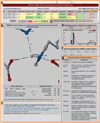
CheckMyMetal (CMM) server: the various components of the results display interface. (a) Summary table with one metal binding site per row. Residue ID, type of metal, and all parameters evaluated are shown for each metal binding site individually. (b) Interactive metal binding site viewer implemented using Jmol. (c) References. (d) Utility to retrieve a model containing alternative metal ion(s). (e) Metal-ligand distribution of the analyzed structure (shown as red columns) and from CSD (shown as blue lines). (f) Shortcuts to retrieve recent jobs submitted from the same client computer. (g) Brief descriptions of the columns listed in the results summary table.
Figure 4
Figure 4
Examples of problematic metal binding sites in crystal structures, as detected by CMM. Metal binding sites are shown before (protein backbone in green) and after re-refinement (backbone in cyan). Electron density maps (2Fo-Fc) are shown in gray mesh with 1.0σ cutoff, with difference maps (Fo-Fc) shown in green and red mesh with 4.0σ cutoff. Only one of the three modeled magnesium ions (A902) is shown for 3lkm, together with the re-refined site with a potassium ion. Another one of the three modeled magnesium ions in 3lkm as replaced with water (A901) is shown in Supplementary Fig. 3. In the 3lrk re-refined structure, only one of the two alternative glycerol molecules is shown for clarity.

Similar articles

Cited by

References

    1. Harding MM, Nowicki MW, Walkinshaw MD. Metals in protein structures: a review of their principal features. Crystallogr Rev. 2010;16:247–302.
    1. Berman HM, et al. The Protein Data Bank. Nucleic Acids Res. 2000;28:235–242. - PMC - PubMed
    1. Pozharski E, Weichenberger CX, Rupp B. Techniques, tools and best practices for ligand electron-density analysis and results from their application to deposited crystal structures. Acta Crystallogr. D. 2013;69:150–167. - PubMed
    1. Chruszcz M, Domagalski M, Osinski T, Wlodawer A, Minor W. Unmet challenges of structural genomics. Curr. Opin. Struct. Biol. 2010;20:587–597. - PMC - PubMed
    1. Zheng H, Chruszcz M, Lasota P, Lebioda L, Minor W. Data mining of metal ion environments present in protein structures. J. Inorg. Biochem. 2008;102:1765–1776. - PMC - PubMed

Publication types

LinkOut - more resources