. 2014 Jan;58(1):69-81.
doi: 10.1007/s10858-013-9806-y. Epub 2013 Dec 20.

Resonance assignments of a membrane protein in phospholipid bilayers by combining multiple strategies of oriented sample solid-state NMR

Affiliations

Resonance assignments of a membrane protein in phospholipid bilayers by combining multiple strategies of oriented sample solid-state NMR

George J Lu et al. J Biomol NMR. 2014 Jan.

Abstract

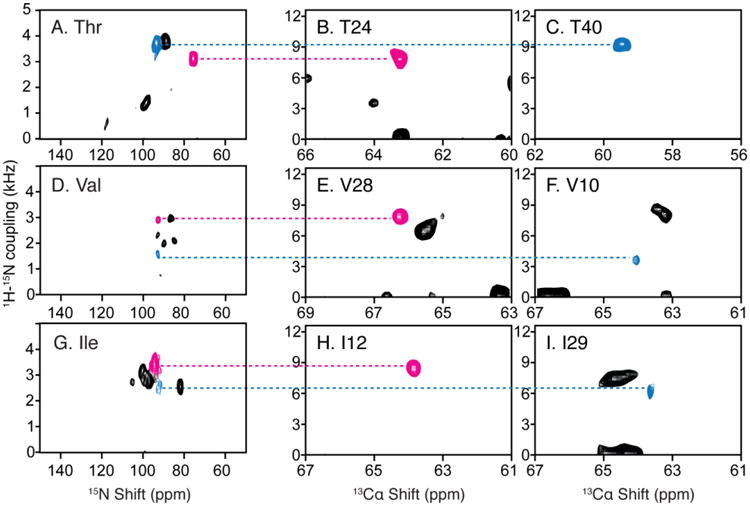
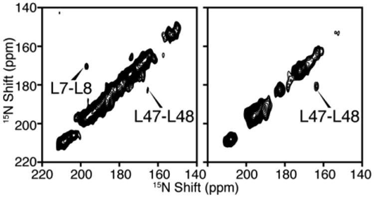
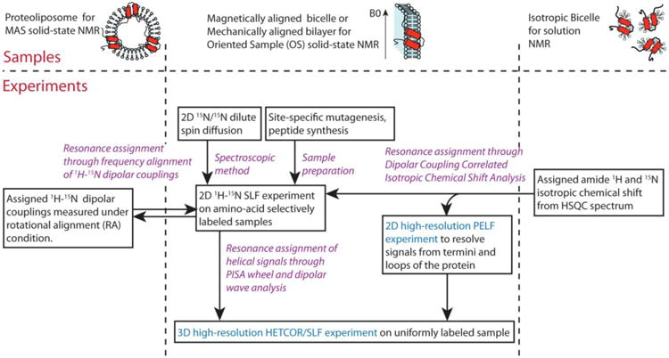
Oriented sample solid-state NMR spectroscopy can be used to determine the three-dimensional structures of membrane proteins in magnetically or mechanically aligned lipid bilayers. The bottleneck for applying this technique to larger and more challenging proteins is making resonance assignments, which is conventionally accomplished through the preparation of multiple selectively isotopically labeled samples and performing an analysis of residues in regular secondary structure based on Polarity Index Slant Angle (PISA) Wheels and Dipolar Waves. Here we report the complete resonance assignment of the full-length mercury transporter, MerF, an 81-residue protein, which is challenging because of overlapping PISA Wheel patterns from its two trans-membrane helices, by using a combination of solid-state NMR techniques that improve the spectral resolution and provide correlations between residues and resonances. These techniques include experiments that take advantage of the improved resolution of the MSHOT4-Pi4/Pi pulse sequence; the transfer of resonance assignments through frequency alignment of heteronuclear dipolar couplings, or through dipolar coupling correlated isotropic chemical shift analysis; (15)N/(15)N dilute spin exchange experiments; and the use of the proton-evolved local field experiment with isotropic shift analysis to assign the irregular terminal and loop regions of the protein, which is the major "blind spot" of the PISA Wheel/Dipolar Wave method.


Figures

Fig. 1
Fig. 1
15N solid-state NMR spectrum of uniformly 15N labeled full-length MerF in q=3.2 14-O-PC/6-O-PC bicelles aligned with their normals perpendicular to the direction of the magnetic field. Line widths of selected resonances are marked in the figure.
Fig. 2
Fig. 2
Two-dimensional SLF and three-dimensional HECTOR/SLF NMR spectra of uniformly and selectively 15N labeled samples of MerF in q=3.2 14-O-PC/6-O-PC magnetically aligned bilayers (q=3.2 bicelles). A. Two-dimensional SLF spectrum of uniformly 15N labeled MerF. B. and C. Two-dimensional planes at the designated 1H chemical shift frequencies from a three-dimensional HECTOR/SLF spectrum. D. Two-dimensional SLF spectrum of selectively 15N leucine labeled MerF. E. and F. Two-dimensional planes at the designated 1H chemical shift frequencies from a three-dimensional HECTOR/SLF spectrum. G-O. Two-dimensional SLF spectrum of other selectively labeled MerF samples. A-C are reprinted from (Lu et al. 2012). D and G-O are from (Howell 2007).
Fig. 3
Fig. 3
Example of the assignment of the isoleucine resonances of MerF by the method of heteronuclear dipolar coupling correlated isotropic chemical shift analysis. A., B., C., and D. are two-dimensional spectra of MerF in q=3.2 14-O-PC/6-O-PC bicelles. A. and B. are “flipped” with the bilayer normal parallel to the field; and C. and D. are “unflipped” with the bilayer normal perpendicular to the field. A. and C. are SLF spectra. B. and D. are HETCOR spectra. The assignment derived from combined DCCICS and PISA wheel analysis are labeled in spectra C and D. Insets in D are extracted from three-dimensional HETCOR/SLF spectrum of the same sample to occupy the missing peaks in two-dimensional spectrum due to selective magnetization transfer.
Fig. 4
Fig. 4
Resonance assignments of the terminal and loop regions of membrane proteins with irregular tertiary structure. PELF spectra of uniformly 15N labeled MerF in flipped (A.) and unflipped (B.) bicelles. C. 1H and 15N isotropic chemical shifts calculated from the spectra in panels A. and B. and three-dimensional HETCOR/SLF spectra. D. Solution HSQC NMR spectrum and the chemical shift of side chains. E. and F. Expansion of the backbone amide resonance region of the spectra in panels C. and D. Those resonances whose assignments can be determined from selectively labeled spectra are labeled in cyan color. In this group, one of the ArgNε peak (magenta color) is an example that DCCICS analysis is crucial for avoiding its mis-assignment as a backbone amide resonance. The remaining resonances that could not be selectively labeled (yellow color) are successfully assigned by DCCICS analysis. A is reprinted from (Lu et al. 2012) after adding resonance assignments.
Fig. 5
Fig. 5
Resonance assignments of residues in OS solid-state NMR spectra utilizing the 1H-15N dipolar couplings measured in RA MAS solid-state NMR experiments. A. D. and G. Two-dimensional SLF spectrum of amino-acid-type selectively 15N labeled MerF in ‘unflipped’ DMPC/DHPC bicelles. B. C. E. F. H. and I. Two-dimensional SLF spectral planes extracted from a three-dimensional HnNCa experiment, of which the third dimension is 15N chemical shift (Lu et al. 2013). The scales during aligning the 1H-15N dipolar couplings are adjusted for the scaling factors of the bicelles vs. liposomes (0.8) and of the alignment perpendicular to the magnetic field vs. rotational alignment parallel to the bilayer normal (0.5).
Fig. 6
Fig. 6
Two-dimensional 15N/15N dilute spin exchange spectra of selectively 15N-Leu labeled MerF in DMPC/DHPC bicelles aligned with their bilayer normal parallel to the magnetic field. A. Spectrum obtained using Mismatched Hartmann-Hahn (MMHH) spin diffusion (Nevzorov 2008). B. Spectrum obtained using Proton-driven Spin Diffusion (PDSD) (Cross et al. 1983).
Fig. 7
Fig. 7
A roadmap for resonance assignments of membrane protein samples in OS solid-state NMR. Purple highlights the five complementary resonance assignment methods. Cyan highlights the experiments with significantly improved resolution by new pulse sequences.

Similar articles

Cited by

References

    1. Banigan J, Gayen A, Traaseth N. Combination of 15N reverse labeling and afterglow spectroscopy for assigning membrane protein spectra by magic-angle-spinning solid-state NMR: application to the multidrug resistance protein EmrE. J Biomol NMR. 2013;55(4):391–399. doi: 10.1007/s10858-013-9724-z. - DOI - PMC - PubMed
    1. Cavanagh J, Fairbrother WJ, Palmer AG, III, Skelton NJ. Protein NMR spectroscopy: principles and practice. Academic Press; San Diego: 1996.
    1. Chekmenev EY, Gor'kov PL, Cross TA, Alaouie AM, Smirnov AI. Flow-Through Lipid Nanotube Arrays for Structure-Function Studies of Membrane Proteins by Solid-State NMR Spectroscopy. Biophys J. 2006;91(8):3076–3084. doi: 10.1529/biophysj.106.085191. - DOI - PMC - PubMed
    1. Cross T, Murray D, Watts A. Helical membrane protein conformations and their environment. Eur Biophys J. 2013:1–25. doi: 10.1007/s00249-013-0925-x. - DOI - PMC - PubMed
    1. Cross TA, Frey MH, Opella SJ. Nitrogen-15 spin exchange in a protein. J Am Chem Soc. 1983;105(25):7471–7473. doi: 10.1021/ja00363a060. - DOI

Publication types

MeSH terms