. 2014 Feb;136(2):021029.
doi: 10.1115/1.4026285.

Analysis of collagen organization in mouse achilles tendon using high-frequency ultrasound imaging

Analysis of collagen organization in mouse achilles tendon using high-frequency ultrasound imaging

Corinne N Riggin et al. J Biomech Eng. 2014 Feb.

Abstract

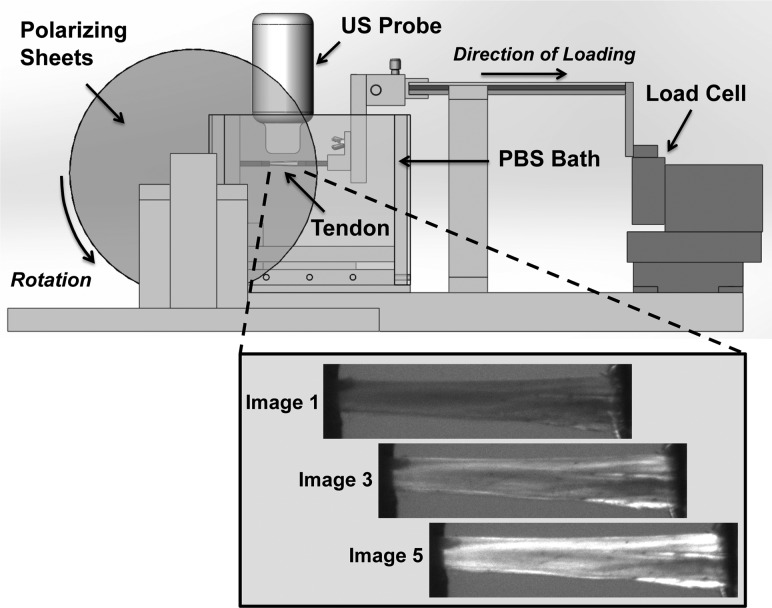
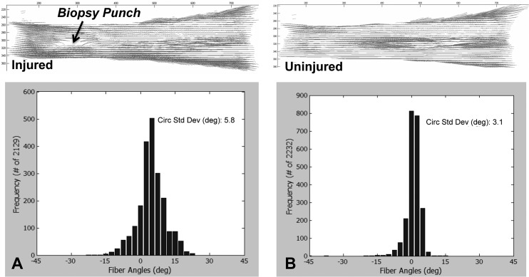
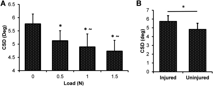
Achilles tendon ruptures are traumatic injuries, and techniques for assessing repair outcomes rely on patient-based measures of pain and function, which do not directly assess tendon healing. Consequently, there is a need for a quantitative, in vivo measure of tendon properties. Therefore, the purpose of this study was to validate ultrasound imaging for evaluating collagen organization in tendons. In this study, we compared our novel, high-frequency ultrasound (HFUS) imaging and analysis method to a standard measure of collagen organization, crossed polarizer (CP) imaging. Eighteen mouse Achilles tendons were harvested and placed into a testing fixture where HFUS and CP imaging could be performed simultaneously in a controlled loading environment. Two experiments were conducted: (1) effect of loading on collagen alignment and (2) effect of an excisional injury on collagen alignment. As expected, it was found that both the HFUS and CP methods could reliably detect an increase in alignment with increasing load, as well as a decrease in alignment with injury. This HFUS method demonstrates that structural measures of collagen organization in tendon can be determined through ultrasound imaging. This experiment also provides a mechanistic evaluation of tissue structure that could potentially be used to develop a targeted approach to aid in rehabilitation or monitor return to activity after tendon injury.


Figures

Fig. 1
Fig. 1
Schematic of setup for capturing CP and HFUS imaging simultaneously. The tendon is secured between two grips in a PBS tank, where the ultrasound probe can be centered over top of the tendon, submerged in the PBS. Two polarizing sheets are located on either side of the tank, with a linear backlight on one side and a camera on the other (not depicted in this image). A custom-built tensiometer is attached to a sliding shaft to allow for load control. Inset figure (below) shows representative images of a tendon as captured by the CP camera through one rotation cycle.
Fig. 2
Fig. 2
(a) HFUS image of Achilles tendon with excisional injury, (b) filtered HFUS image, (c) hyper- and hypoechoic regions that create the banded pattern that is quantified as the CSD of the fiber orientations
Fig. 3
Fig. 3
Alignment maps and histograms depicting the distribution of localized fiber directions throughout the tendon that were produced through CP analysis for (a) injured and (b) uninjured specimens
Fig. 4
Fig. 4
(a) Changes in CSD in response to load for the HFUS images. There is a significant decrease in CSD (increase in organization) with increased loading. Statistics for each load compared to 0 N (*p < 0.05) and to 0.5 N (∼p < 0.05). (b) CSD measures for HFUS images before and after a biopsy punch excisional injury. There is a significant increase in CSD (decrease in organization) with injury (*p < 0.05).
Fig. 5
Fig. 5
(a) Changes in CSD in response to load for the CP images. There is a significant decrease in CSD (increase in organization) with increased loading. Statistics for each load compared to 0 N (*p < 0.05) and to 0.5 N (∼p < 0.05). (b) CSD measures for CP images before and after a biopsy punch excisional injury. There is an increase in CSD (decrease in organization) with injury (*p < 0.05).

Similar articles

Cited by

References

    1. Suchak, A. A. , Bostick, G. , Reid, D. , Blitz, S. , and Jomha, N. , 2005, “The Incidence of Achilles Tendon Ruptures in Edmonton, Canada,” Foot Ankle Int., 26(11), pp. 932–936. - PubMed
    1. Houshian, S. , Tscherning, T. , and Riegels-Nielsen, P. , 1998, “The Epidemiology of Achilles Tendon Rupture in a Danish County,” Injury, 29(9), pp. 651–654.10.1016/S0020-1383(98)00147-8 - DOI - PubMed
    1. Moller, A. , Astron, M. , and Westlin, N. , 1996, “Increasing Incidence of Achilles Tendon Rupture,” Acta Orthop. Scand., 67(5), pp. 479–481.10.3109/17453679608996672 - DOI - PubMed
    1. Lin, T. W. , Cardenas, L. , and Soslowsky, L. J. , 2004, “Biomechanics of Tendon Injury and Repair,” J. Biomech., 37(6), pp. 865–877.10.1016/j.jbiomech.2003.11.005 - DOI - PubMed
    1. Saxena, A. , Ewen, B. , and Maffulli, N. , 2011, “Rehabilitation of the Operated Achilles Tendon: Parameters for Predicting Return to Activity,” J. Foot Ankle Surg., 50(1), pp. 37–40.10.1053/j.jfas.2010.10.008 - DOI - PubMed

Publication types