. 2014 Feb 7;289(6):3749-62.
doi: 10.1074/jbc.M113.512756. Epub 2013 Dec 19.

Characterization and structure of the vaccinia virus NF-κB antagonist A46

Affiliations

Characterization and structure of the vaccinia virus NF-κB antagonist A46

Sofiya Fedosyuk et al. J Biol Chem. .

Abstract

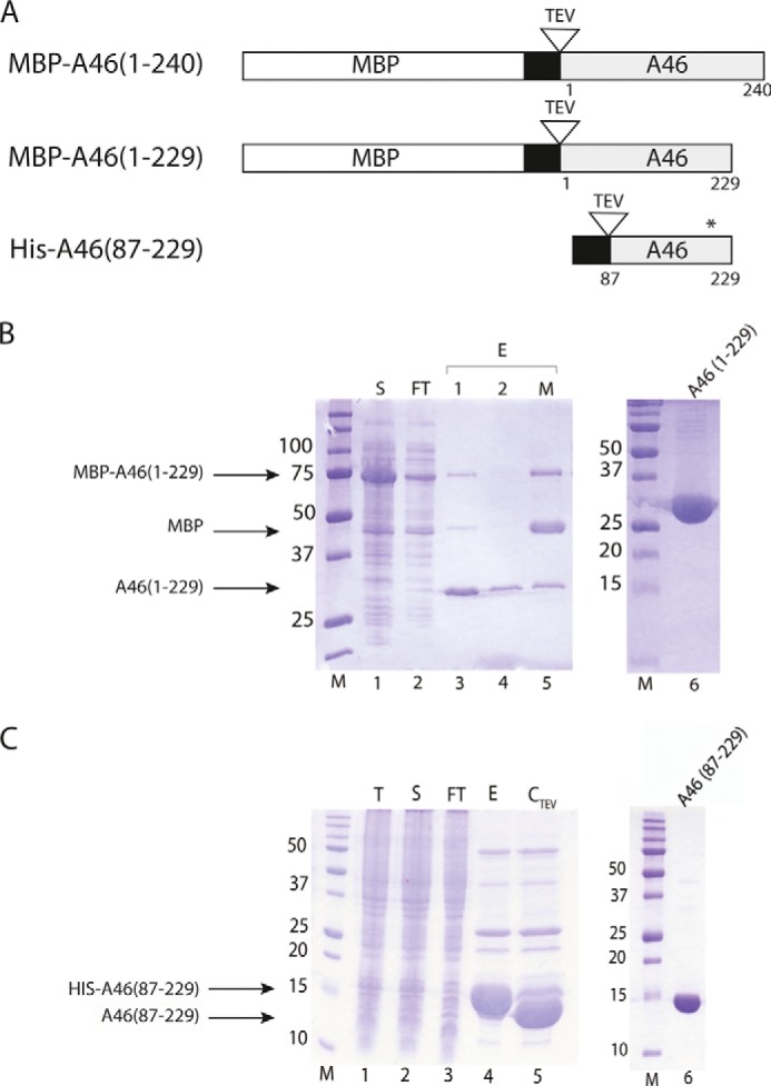
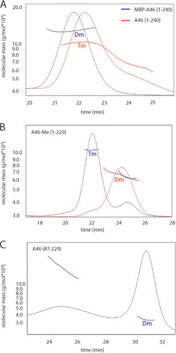
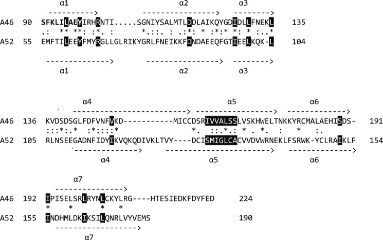
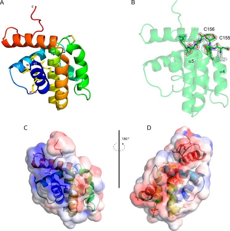
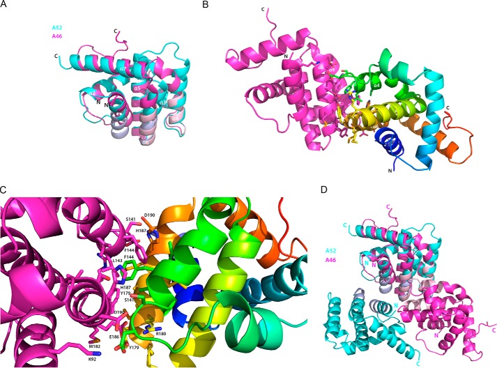
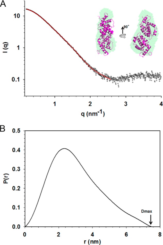
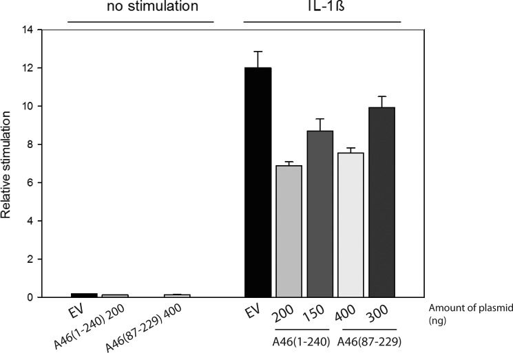
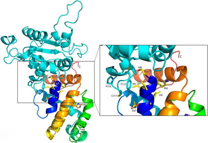
Successful vaccinia virus (VACV) replication in the host requires expression of viral proteins that interfere with host immunity, such as antagonists of the activation of the proinflammatory transcription factor NF-κB. Two such VACV proteins are A46 and A52. A46 interacts with the Toll-like receptor/interleukin-1R (TIR) domain of Toll-like receptors and intracellular adaptors such as MAL (MyD88 adapter-like), TRAM (TIR domain-containing adapter-inducing interferon-β (TRIF)-related adaptor molecule), TRIF, and MyD88, whereas A52 binds to the downstream signaling components TRAF6 and IRAK2. Here, we characterize A46 biochemically, determine by microscale thermophoresis binding constants for the interaction of A46 with the TIR domains of MyD88 and MAL, and present the 2.0 Å resolution crystal structure of A46 residues 87-229. Full-length A46 behaves as a tetramer; variants lacking the N-terminal 80 residues are dimeric. Nevertheless, both bind to the Toll-like receptor domains of MAL and MyD88 with KD values in the low μm range. Like A52, A46 also shows a Bcl-2-like fold but with biologically relevant differences from that of A52. Thus, A46 uses helices α4 and α6 to dimerize, compared with the α1-α6 face used by A52 and other Bcl-2 like VACV proteins. Furthermore, the loop between A46 helices α4-α5 is flexible and shorter than in A52; there is also evidence for an intramolecular disulfide bridge between consecutive cysteine residues. We used molecular docking to propose how A46 interacts with the BB loop of the TRAM TIR domain. Comparisons of A46 and A52 exemplify how subtle changes in viral proteins with the same fold lead to crucial differences in biological activity.

Keywords: Bcl-2 Family Proteins; Inflammation; MyD88; Protein-Protein Interactions; Viral Immunology.


Figures

FIGURE 1.
FIGURE 1.
Expression and purification of A46 variants. A, schematic representation of A46 variants used in this work. The MBP tag is indicated by the open bar, the His tag is indicated by the black bar, the A46 is indicated by the gray bar, and the TEV site is indicated by an open triangle. The position of the modified Cys-205 residue is marked by an asterisk. B, SDS-PAGE analysis of the purification of MBP-A46-(1–229). Samples of total protein from each of the indicated fractions were applied to gels containing 12.5% (except for lane 6, 15%) acrylamide. Lane 1, soluble protein loaded onto amylose column (S); lane 2, amylose column flow-through (FT); lanes 3 and 4, eluate from the amylose column after TEV cleavage (E1, E2); lane 5, eluate from the amylose column after TEV cleavage in the presence of maltose. Lane 6, 15 μg of concentrated pooled fraction after SEC (note 15% acrylamide percentage). M, molecular mass markers. C, SDS-PAGE analysis on gels containing 15% acrylamide of the purification of His-A46-(87–229). Lane 1, crude cell extract (T); lane 2, total soluble protein in crude cell extract loaded on to Ni-NTA beads (S); lane 3, flow-through from Ni-NTA beads (FT); lane 4, eluate from Ni-NTA beads with imidazole (E); lane 5, eluate after incubation with TEV protease (CTEV); lane 6, 7.5 μg concentrated pooled fraction after SEC. Proteins were visualized with Coomassie Brilliant Blue R250. Numbers on the left indicate apparent molecular sizes (in kDa).
FIGURE 2.
FIGURE 2.
Examination of the solution oligomeric states of indicated A46 variants by SEC-multiangle laser light scattering. A, wild-type A46 behaves as a tetramer (Tm), whereas MBP tagged full-length A46 behaves a dimer (Dm). B, methylated A46-(1–229) was separated into tetramer and dimer populations by size-exclusion chromatography, and the resulting fractions were subjected to static light scattering. C, The N- and C-terminally truncated A46-(87–229) variant that was successfully crystallized behaves as a dimer on static light scattering. 100–200 μg of the indicated proteins was applied to a Superdex 10/300 column, and the light scattering was measured using a miniDAWN Tristar light scattering instrument. Molecular masses were calculated directly from the light scattering measurements.
FIGURE 3.
FIGURE 3.
Analysis of A46 interactions with microscale thermophoresis. Unlabeled full-length A46C205 (closed circles, 1.8 nm to 30 μm) or A46-(87–229) (open circles, 5.2 nm to 170 μm) were titrated into a fixed concentration of labeled TIR/MyD88 (30–70 nm). Both A46 variants demonstrated similar binding properties with apparent KD values of 0.5 and about 1.0 μm, respectively. Values are an average of the two measurements presented in Table 3.
FIGURE 4.
FIGURE 4.
Amino acid alignment of A46 and A52 showing secondary structure alignments. A sequence alignment was first generated using the T-coffee algorithm (59) and then modified by hand. Residues identified by Franklin and Khan (27) as forming the hydrophobic core of vaccinia proteins with a Bcl-2 fold are shown in reverse, as are the conserved arginine and aspartic acid residues in α1 and α2, respectively. Residues 90–98 that make up part of the VIPER peptide are shown in bold.
FIGURE 5.
FIGURE 5.
Overall structure of A46. A, view of the overall structure of A46 colored as a spectrum from the N terminus to the C terminus. The side chains of the nine residues of the 11-mer VIPER peptide visible in the structure are shown as sticks as are those of Cys-155 and Cys-156. Carbon atoms are colored in light yellow, oxygen atoms are in red, nitrogen atoms are in blue, and sulfur atoms are in dark yellow. B, 2FoFc electron density map contoured at 1.2δ of the minor confirmation of C156 observed in molecule A, illustrating the disulfide bridge to C155. C and D, electrostatic surface potentials of A46, blue being electropositive and red being electronegative. In C the orientation of the molecule is that in A, in D it is rotated 180°. Drawings were made with PyMOL (60) and APBS (61).
FIGURE 6.
FIGURE 6.
Comparison of the structures of A46 and A52. A, superimposition of A46 (magenta) to residues 54–189 of A52 (cyan). Faces involved in dimer formation are shown in light pink (A46) and light blue (A52). B, the A46 dimer. Molecule C of A46 (magenta) is in the same orientation as in Fig. 6A, and molecule D is colored as a spectrum from the N terminus to the C terminus. Residues involved in dimer formation are shown as sticks. C, enlarged view of the residues in the A46 dimer interface shown in A, rotated 45° on the x axis to allow better visualization of the side chains. D, as in A, except that the second monomer is included for each protein. Drawings were made as in Fig. 5.
FIGURE 7.
FIGURE 7.
Analysis of scattering data of A46. A, average ab initio bead model of A46 (green) fits the dimer from crystal structure (magenta). The experimental SAXS data are represented as an open sphere, whereas the ab initio fit is in red. The data were fit at resolution of about 25 Å. B, distance distribution analysis calculated using SAXS data.
FIGURE 8.
FIGURE 8.
Inhibition of IL-1β stimulation of NF-κβ transcription by A46. HEK293T cells were transfected with the indicated amounts of plasmids. After 40 h of incubation, the cells were stimulated with 0.32 ng/ml of IL-1β and incubated for a further 6 h. NF-κβ reporter gene activity was then measured. Data are expressed as the relative stimulation from a representative experiment from a minimum three separate experiments, each performed in triplicate. EV, empty vector
FIGURE 9.
FIGURE 9.
Docking model of A46 with TRAM. The coordinates of A46 and the model of TRAM generated by (29) were submitted to the ClusPro protein-protein docking server (62). One docking result that showed an interaction of the VIPER peptide with the BB-loop of TRAM was chosen and visualized. A46 is colored as a spectrum from the N terminus to the C terminus, the VIPER residues are shown as sticks (carbon atoms are colored in light yellow, oxygen atoms are in red, and nitrogen atoms are in blue) as are residues Phe-144 and His-187 to mark the 4–6 dimer interface. TRAM is colored in cyan, with the residues of the BB loop are shown in sticks. The inset shows an enlargement of the VIPER sequence of A46 and the BB loop of TRAM.

Similar articles

Cited by

References

    1. Medzhitov R., Janeway C., Jr. (2000) Innate immunity. N. Engl. J. Med. 343, 338–344 - PubMed
    1. Aoshi T., Koyama S., Kobiyama K., Akira S., Ishii K. J. (2011) Innate and adaptive immune responses to viral infection and vaccination. Curr. Opin. Virol. 1, 226–232 - PubMed
    1. O'Neill L. A. (2006) How Toll-like receptors signal. What we know and what we don't know. Curr. Opin. Immunol. 18, 3–9 - PubMed
    1. Bowie A. G., Unterholzner L. (2008) Viral evasion and subversion of pattern-recognition receptor signalling. Nat. Rev. Immunol. 8, 911–922 - PMC - PubMed
    1. Kotwal G. J., Hatch S., Marshall W. L. (2012) Viral infection. An evolving insight into the signal transduction pathways responsible for the innate immune response. Adv. Virol. 2012, 131457. - PMC - PubMed

Publication types

Associated data