Fenretinide induces ubiquitin-dependent proteasomal degradation of stearoyl-CoA desaturase in human retinal pigment epithelial cells
- PMID: 24357007
- PMCID: PMC3999186
- DOI: 10.1002/jcp.24527
Fenretinide induces ubiquitin-dependent proteasomal degradation of stearoyl-CoA desaturase in human retinal pigment epithelial cells
Abstract
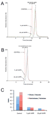
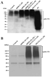
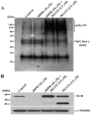
Stearoyl-CoA desaturase (SCD, SCD1), an endoplasmic reticulum (ER) resident protein and a rate-limiting enzyme in monounsaturated fatty acid biosynthesis, regulates cellular functions by controlling the ratio of saturated to monounsaturated fatty acids. Increase in SCD expression is strongly implicated in the proliferation and survival of cancer cells, whereas its decrease is known to impair proliferation, induce apoptosis, and restore insulin sensitivity. We examined whether fenretinide, (N-(4-hydroxyphenyl)retinamide, 4HPR), which induces apoptosis in cancer cells and recently shown to improve insulin sensitivity, can modulate the expression of SCD. We observed that fenretinide decreased SCD protein and enzymatic activity in the ARPE-19 human retinal pigment epithelial cell line. Increased expression of BiP/GRP78, ATF4, and GADD153 implicated ER stress. Tunicamycin and thapsigargin, compounds known to induce ER stress, also decreased the SCD protein. This decrease was completely blocked by the proteasome inhibitor MG132. In addition, PYR41, an inhibitor of ubiquitin activating enzyme E1, blocked the fenretinide-mediated decrease in SCD. Immunoprecipitation analysis using anti-ubiquitin and anti-SCD antibodies and the blocking of SCD loss by PYR41 inhibition of ubiquitination further corroborate that fenretinide mediates the degradation of SCD in human RPE cells via the ubiquitin-proteasome dependent pathway. Therefore, the effect of fenretinide on SCD should be considered in its potential therapeutic role against cancer, type-2 diabetes, and retinal diseases.
© 2013 Wiley Periodicals, Inc.
Figures


Similar articles
-
Pretreatment of human retinal pigment epithelial cells with sterculic acid forestalls fenretinide-induced apoptosis.Sci Rep. 2022 Dec 23;12(1):22442. doi: 10.1038/s41598-022-26383-9. Sci Rep. 2022. PMID: 36575190 Free PMC article.
-
MicroRNA expression in human retinal pigment epithelial (ARPE-19) cells: increased expression of microRNA-9 by N-(4-hydroxyphenyl)retinamide.Mol Vis. 2010 Aug 4;16:1475-86. Mol Vis. 2010. PMID: 20806079 Free PMC article.
-
Modulation of palmitate-induced endoplasmic reticulum stress and apoptosis in pancreatic β-cells by stearoyl-CoA desaturase and Elovl6.Am J Physiol Endocrinol Metab. 2011 Apr;300(4):E640-9. doi: 10.1152/ajpendo.00544.2010. Epub 2011 Jan 25. Am J Physiol Endocrinol Metab. 2011. PMID: 21266672
-
Regulation of stearoyl-CoA desaturase by polyunsaturated fatty acids and cholesterol.J Lipid Res. 1999 Sep;40(9):1549-58. J Lipid Res. 1999. PMID: 10484602 Review.
-
Stearoyl-CoA desaturase-1 and adaptive stress signaling.Biochim Biophys Acta. 2016 Nov;1861(11):1719-1726. doi: 10.1016/j.bbalip.2016.08.009. Epub 2016 Aug 24. Biochim Biophys Acta. 2016. PMID: 27550503 Review.
Cited by
-
RUNX2 interacts with SCD1 and activates Wnt/β-catenin signaling pathway to promote the progression of clear cell renal cell carcinoma.Cancer Med. 2023 Mar;12(5):5764-5780. doi: 10.1002/cam4.5326. Epub 2022 Oct 6. Cancer Med. 2023. PMID: 36200301 Free PMC article.
-
Paeoniflorin inhibits human glioma cells via STAT3 degradation by the ubiquitin-proteasome pathway.Drug Des Devel Ther. 2015 Oct 13;9:5611-22. doi: 10.2147/DDDT.S93912. eCollection 2015. Drug Des Devel Ther. 2015. PMID: 26508835 Free PMC article.
-
Accumulation of cholesterol and increased demand for zinc in serum-deprived RPE cells.Mol Vis. 2016 Dec 10;22:1387-1404. eCollection 2016. Mol Vis. 2016. PMID: 28003730 Free PMC article.
-
Phyllanthus Niruri L. Exerts Protective Effects Against the Calcium Oxalate-Induced Renal Injury via Ellgic Acid.Front Pharmacol. 2022 Jun 16;13:891788. doi: 10.3389/fphar.2022.891788. eCollection 2022. Front Pharmacol. 2022. PMID: 36034880 Free PMC article.
-
Pretreatment of human retinal pigment epithelial cells with sterculic acid forestalls fenretinide-induced apoptosis.Sci Rep. 2022 Dec 23;12(1):22442. doi: 10.1038/s41598-022-26383-9. Sci Rep. 2022. PMID: 36575190 Free PMC article.
References
-
- Acharya P, Engel JC, Correia MA. Hepatic CYP3A suppression by high concentrations of proteasomal inhibitors: a consequence of endoplasmic reticulum (ER) stress induction, activation of RNA-dependent protein kinase-like ER-bound eukaryotic initiation factor 2alpha (eIF2alpha)-kinase (PERK) and general control nonderepressible-2 eIF2alpha kinase (GCN2), and global translational shutoff. Mol Pharmacol. 2009;76:503–515. - PMC - PubMed
-
- Camerini T, Mariani L, De Palo G, Marubini E, Di Mauro MG, Decensi A, Costa A, Veronesi U. Safety of the synthetic retinoid fenretinide: long-term results from a controlled clinical trial for the prevention of contralateral breast cancer. Journal of clinical oncology : official journal of the American Society of Clinical Oncology. 2001;19:1664–1670. - PubMed
-
- Chen C, Sun X, Ran Q, Wilkinson KD, Murphy TJ, Simons JW, Dong JT. Ubiquitin-proteasome degradation of KLF5 transcription factor in cancer and untransformed epithelial cells. Oncogene. 2005;24:3319–3327. - PubMed
Publication types
MeSH terms
Substances
Grants and funding
LinkOut - more resources
Full Text Sources
Other Literature Sources
Research Materials
Miscellaneous

