

Effects of dried plum supplementation on bone metabolism in adult C57BL/6 male mice
- PMID: 24357047
- PMCID: PMC3950615
- DOI: 10.1007/s00223-013-9819-2
Effects of dried plum supplementation on bone metabolism in adult C57BL/6 male mice
Abstract
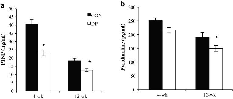
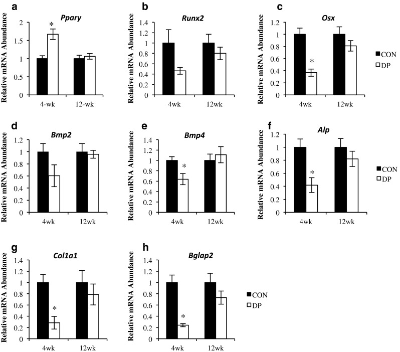
Dietary supplementation of dried plum (DP) prevents bone loss and restores bone mass in osteopenic animal models. This study was designed to determine the effects of DP supplementation on bone metabolic activity over time using adult (6-month-old) male C57BL/6 mice (n = 40) receiving control (CON = AIN93 M) or CON+DP 25 % (w/w) diets for 4 or 12 weeks. After 4 weeks of treatment, animals consuming the DP diet had a higher whole-body bone mineral density, vertebral trabecular bone volume (BV/TV), and femoral cortical thickness compared to the CON animals. In the distal metaphysis of the femur, BV/TV was increased in the DP-treated animals, but only after 12 weeks. Bone histomorphometric analyses revealed that DP decreased osteoblast surface (67 %) and osteoclast surface (62 %) at 4 weeks, but these surfaces normalized to the CON animals by 12 weeks. Coincident with these changes, the mineralizing surface (MS/BS) and cancellous bone formation rate (BFR/BS) were reduced at 4 weeks in the DP group compared to the CON, but by 12 weeks of DP supplementation, BFR/BS (~twofold) and MS/BS (~1.7-fold) tended to be increased (p < 0.10). The relative abundance of RNA for key regulators of osteoblast and osteoclast differentiation and indicators of osteoblast activity were reduced in the DP group at 4 weeks with no difference between groups at 12 weeks. These results indicate that supplementing the diet with DP initially suppressed cancellous bone turnover, but a biphasic response occurs over time, resulting in a positive effect on bone mass and structure.
Figures


Similar articles
-
A comparative study of the bone metabolic response to dried plum supplementation and PTH treatment in adult, osteopenic ovariectomized rat.Bone. 2014 Jan;58:151-9. doi: 10.1016/j.bone.2013.10.005. Epub 2013 Oct 11. Bone. 2014. PMID: 24125756
-
Energy-restricted diet benefits body composition but degrades bone integrity in middle-aged obese female rats.Nutr Res. 2013 Aug;33(8):668-76. doi: 10.1016/j.nutres.2013.05.008. Epub 2013 Jun 24. Nutr Res. 2013. PMID: 23890357
-
Comparison of dried plum supplementation and intermittent PTH in restoring bone in osteopenic orchidectomized rats.Osteoporos Int. 2007 Jul;18(7):931-42. doi: 10.1007/s00198-007-0335-y. Epub 2007 Feb 15. Osteoporos Int. 2007. PMID: 17554580
-
Dietary dried plum increases bone mass, suppresses proinflammatory cytokines and promotes attainment of peak bone mass in male mice.J Nutr Biochem. 2016 Aug;34:73-82. doi: 10.1016/j.jnutbio.2016.04.007. Epub 2016 May 10. J Nutr Biochem. 2016. PMID: 27239754 Free PMC article.
-
Viewpoint: dried plum, an emerging functional food that may effectively improve bone health.Ageing Res Rev. 2009 Apr;8(2):122-7. doi: 10.1016/j.arr.2009.01.002. Ageing Res Rev. 2009. PMID: 19274852 Review.
Cited by
-
The Role of Prunes in Modulating Inflammatory Pathways to Improve Bone Health in Postmenopausal Women.Adv Nutr. 2022 Oct 2;13(5):1476-1492. doi: 10.1093/advances/nmab162. Adv Nutr. 2022. PMID: 34978320 Free PMC article. Review.
-
Dried Plum Polyphenolic Extract Combined with Vitamin K and Potassium Restores Trabecular and Cortical Bone in Osteopenic Model of Postmenopausal Bone Loss.J Funct Foods. 2018 Mar;42:262-270. doi: 10.1016/j.jff.2017.12.057. Epub 2018 Jan 30. J Funct Foods. 2018. PMID: 30319713 Free PMC article.
-
Glucocorticoid-induced osteoporosis is prevented by dietary prune in female mice.Front Cell Dev Biol. 2024 Feb 5;11:1324649. doi: 10.3389/fcell.2023.1324649. eCollection 2023. Front Cell Dev Biol. 2024. PMID: 38375074 Free PMC article.
-
Osteoclast Differentiation is Downregulated by Select Polyphenolic Fractions from Dried Plum via Suppression of MAPKs and Nfatc1 in Mouse C57BL/6 Primary Bone Marrow Cells.Curr Dev Nutr. 2017 Sep 6;1(10):e000406. doi: 10.3945/cdn.117.000406. eCollection 2017 Oct. Curr Dev Nutr. 2017. PMID: 29955675 Free PMC article.
-
Freeze-Dried Watermelon Supplementation Has Modest Effects on Bone and Lipid Parameters of Ovariectomized Mice.Prev Nutr Food Sci. 2020 Mar 31;25(1):41-49. doi: 10.3746/pnf.2020.25.1.41. Prev Nutr Food Sci. 2020. PMID: 32292754 Free PMC article.
References
-
- Surgeon General (2004) Bone health and osteoporosis: a report of the surgeon general. http://www.surgeongeneral.gov/library/reports/bonehealth/ - PubMed
-
- Kiel DP, Rosen CJ, Dempster D. Age-related bone loss. In: Rosen CJ, editor. Primer on the metabolic bone diseases and disorders of mineral metabolism. Washington, DC: American Society of Bone and Mineral Research; 2008. pp. 98–101.
Publication types
MeSH terms
Substances
Grants and funding
LinkOut - more resources
Full Text Sources
Other Literature Sources
