. 2014 Apr;243(4):509-26.
doi: 10.1002/dvdy.24092. Epub 2013 Dec 19.

The Role of Sdf-1α signaling in Xenopus laevis somite morphogenesis

Affiliations

The Role of Sdf-1α signaling in Xenopus laevis somite morphogenesis

Marisa A Leal et al. Dev Dyn. 2014 Apr.

Abstract

Background: Stromal derived factor-1α (sdf-1α), a chemoattractant chemokine, plays a major role in tumor growth, angiogenesis, metastasis, and in embryogenesis. The sdf-1α signaling pathway has also been shown to be important for somite rotation in zebrafish (Hollway et al., 2007). Given the known similarities and differences between zebrafish and Xenopus laevis somitogenesis, we sought to determine whether the role of sdf-1α is conserved in Xenopus laevis.

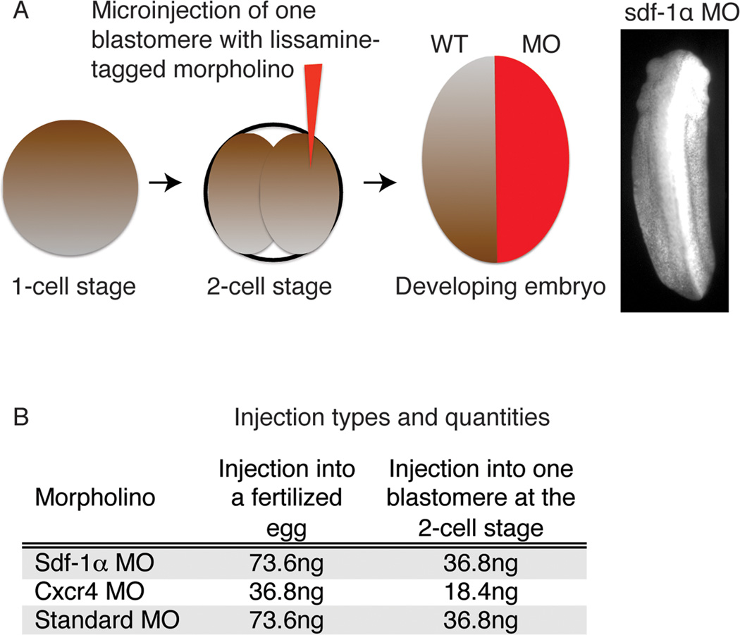
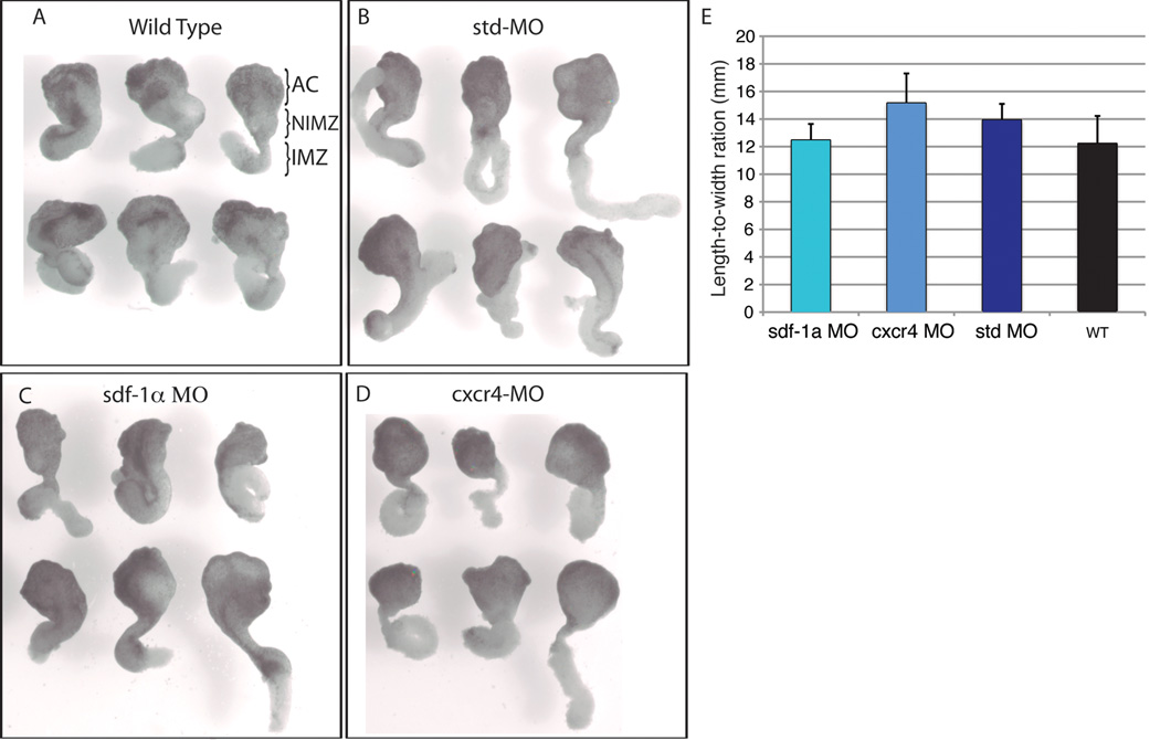
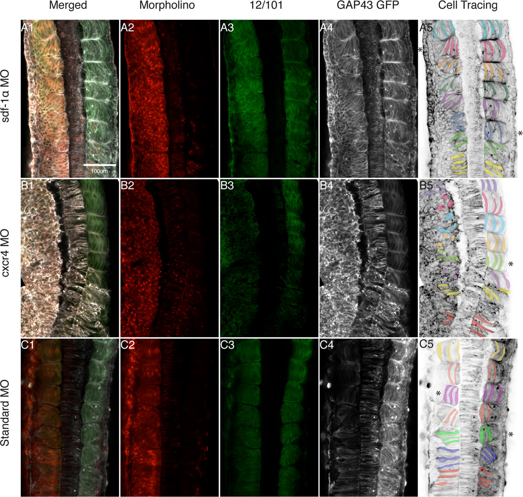
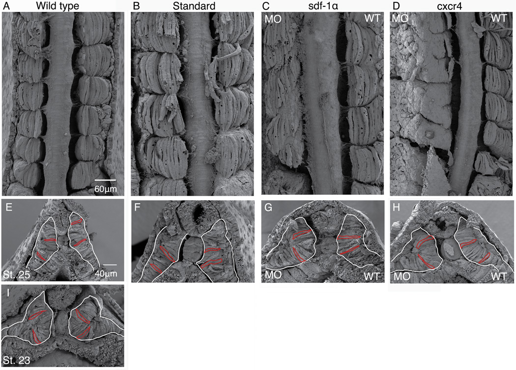
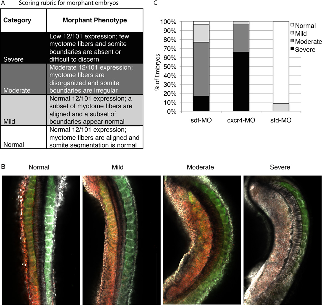
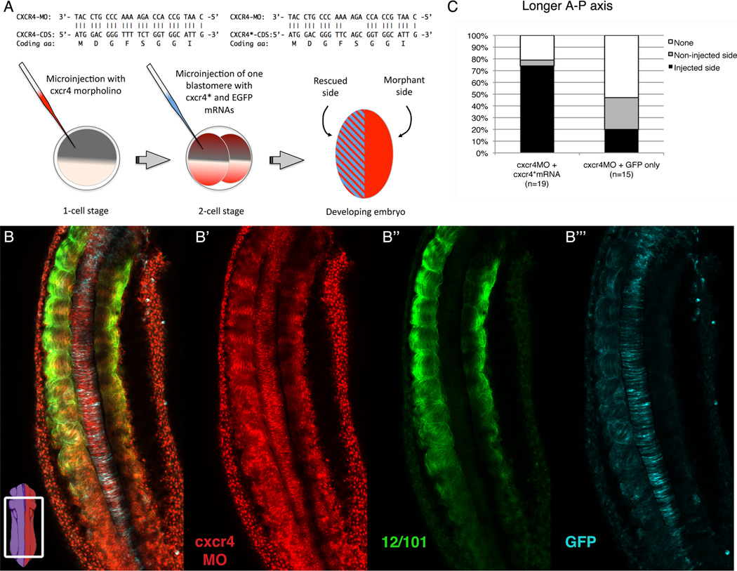
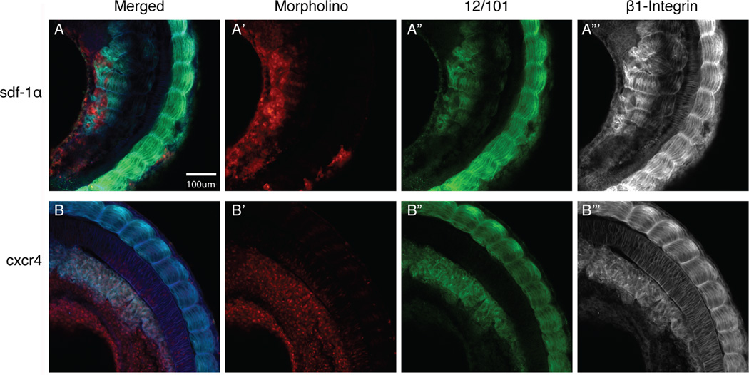
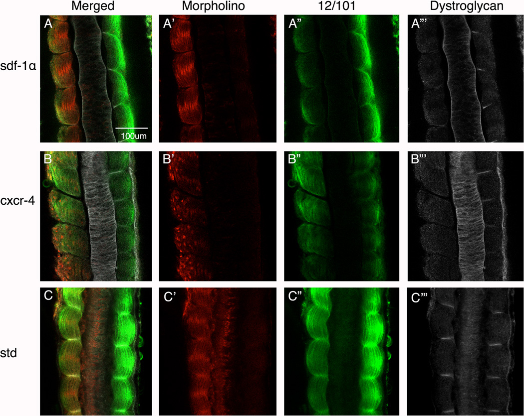
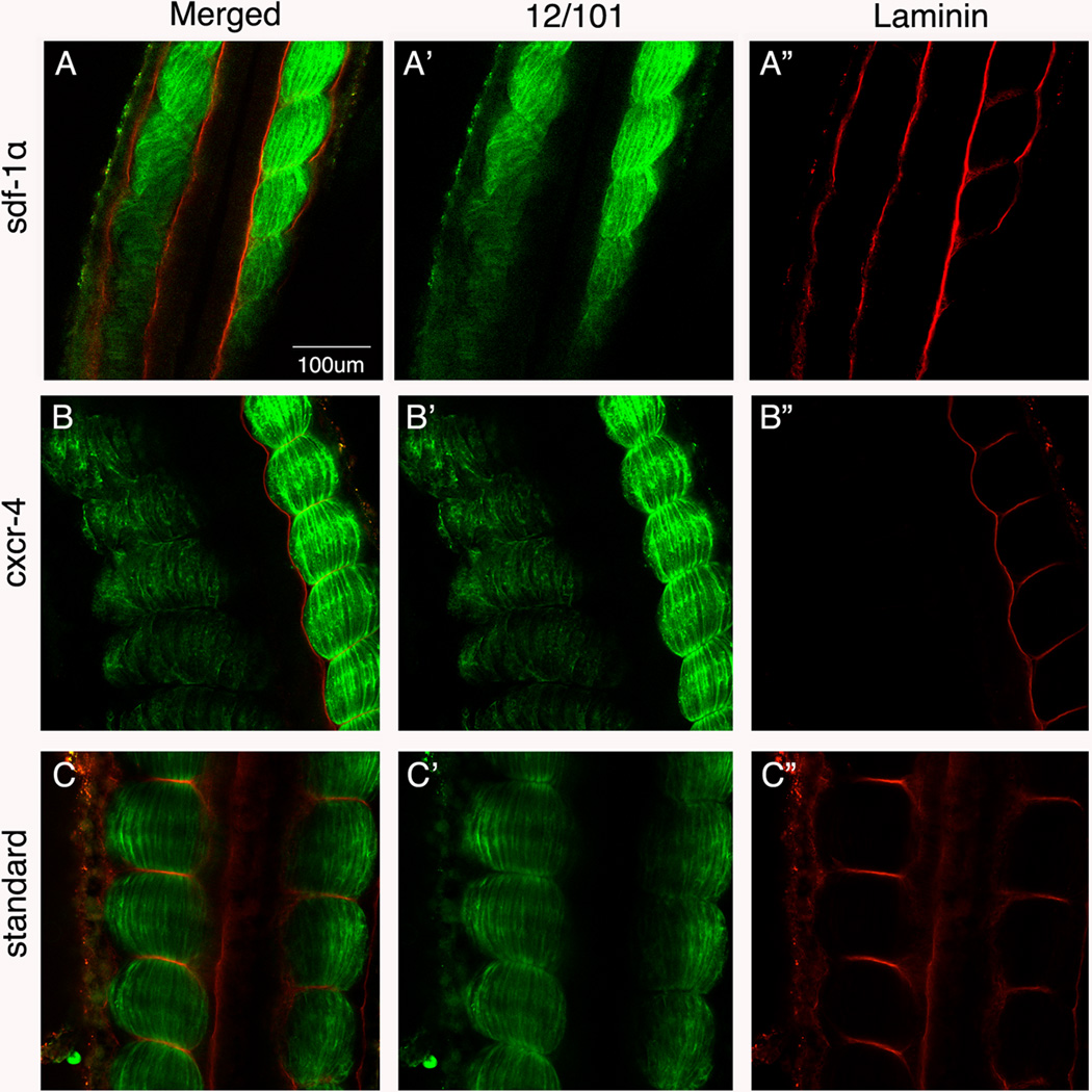
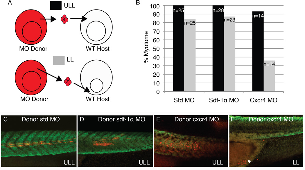
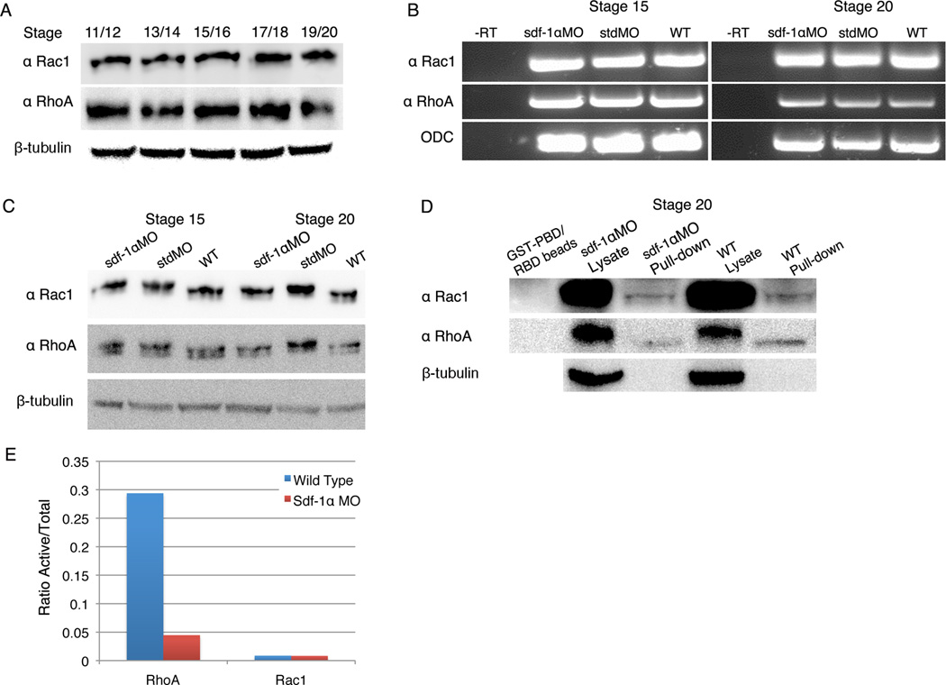
Results: Using a morpholino approach, we demonstrate that knockdown of sdf-1α or its receptor, cxcr4, leads to a significant disruption in somite rotation and myotome alignment. We further show that depletion of sdf-1α or cxcr4 leads to the near absence of β-dystroglycan and laminin expression at the intersomitic boundaries. Finally, knockdown of sdf-1α decreases the level of activated RhoA, a small GTPase known to regulate cell shape and movement.

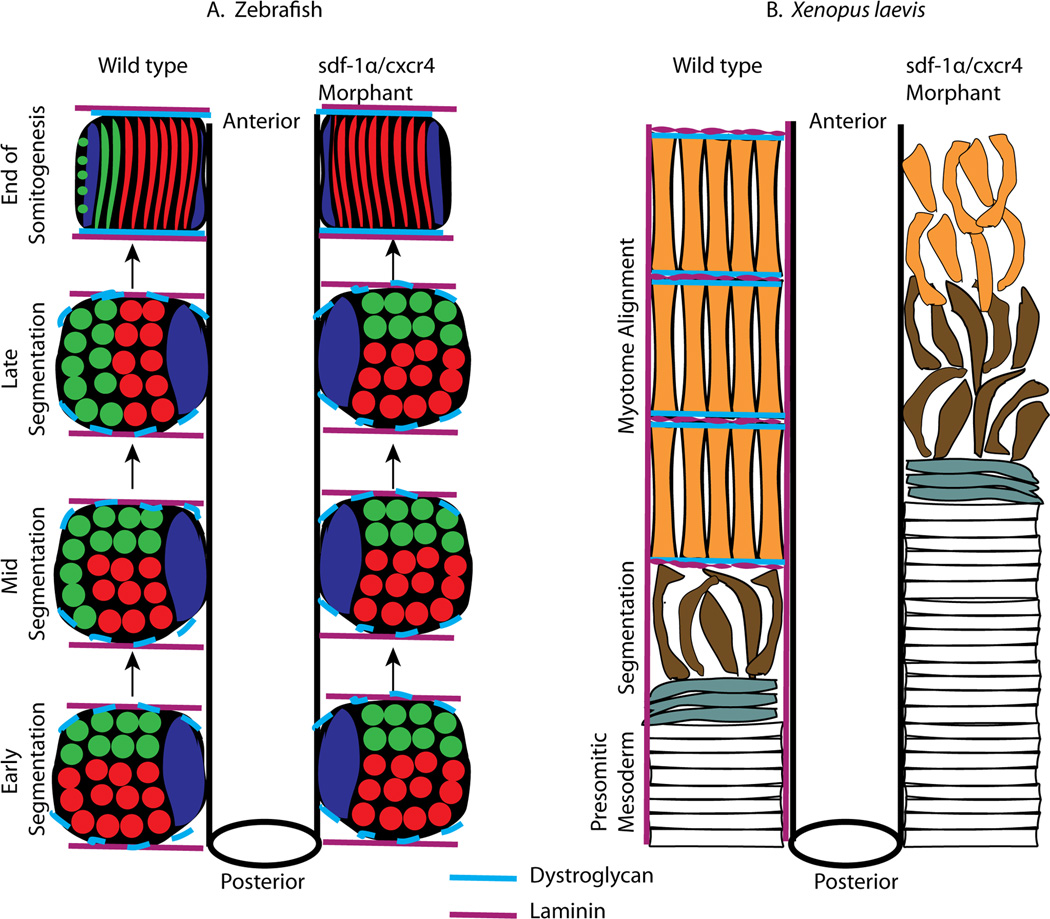
Conclusion: Our results show that sdf-1α signaling regulates somite cell migration, rotation, and myotome alignment by directly or indirectly regulating dystroglycan expression and RhoA activation. These findings support the conservation of sdf-1α signaling in vertebrate somite morphogenesis; however, the precise mechanism by which this signaling pathway influences somite morphogenesis is different between the fish and the frog.

Keywords: RhoA; Xenopus laevis; cxcr4; morphogenesis; muscle; sdf-1α; somite; β-dystroglycan.


Figures

Figure 1
Figure 1. Experimental approach to the morpholino knockdown of sdf-1α and cxcr4
(A) A schematic of the experiment in which one blastomere at the 2-cell stage is injected with a specific morpholino (MO) such that only half of the embryo is morphant. The embryo is then allowed to develop to tailbud stages and then fixed and analyzed. (B) A table indicating the specific amounts of MO injected into the fertilized egg or one blastomere at the two-cell stage.
Figure 2
Figure 2. Time-lapse imaging of sdf-1α and cxcr4 half-morphant embryos
Live time-lapse images of wild type embryos (A–E), standard half-morphants (F–J), sdf-1α half-morphants (K–O), and cxcr4 half-morphants (P–S) as they proceed from gastrulation to the formation of early stage tadpoles (stages 26–28). Scale bar in (A) applies to all frames.
Figure 3
Figure 3. Sdf-1α and cxcr4 are not required for convergent extension
Keller sandwiches made from stage 10 wild type (A), standard MO (B), sdf-1α MO (C), and cxcr4 MO (D) embryos undergo the characteristic convergent extension to form a long and narrow array of cells. (E) Length-to-width ratio of Keller sandwiches reveal no significant differences between explants made from sdf-1α and cxcr4 morphant tissue compared to the controls. Statistical analysis was carried out by using the Student’s t test. Error bars indicate standard error. (AC) Animal Cap. (NIMZ) Non-involuting marginal zone. (IMZ) Involuting marginal zone.
Figure 4
Figure 4. Morpholino depletion of sdf-1α and cxcr4 cause a disruption in somite morphogenesis
Montages of 20X dorsal scans of stage 26 sdf-1α (A.1), cxcr4 (B.1) and standard (C.1) half-morphants highlight the morphologies of cells in the paraxial mesoderm. (A.2–C.2) The distribution of the MO along one-half of the embryo is indicated by lissamine fluorescence. (A.3–C.3) Whole-mount immunocytochemistry with the muscle-specific antibody, 12/101. (A.4–C.4) Black and white images of GAP43 GFP expression. (A.5–C.5) Inverted images of the GAP43 GFP expression with pseudo-coloring of cells to highlight a subset of cell shapes within the paraxial mesoderm. Asterisk indicates the first fully rotated somite. Scale bar in (A1) applies to all frames. Anterior is at the top.
Figure 5
Figure 5. Scanning electron micrographs reveal cell shapes in sdf-1α and cxcr4 morphant embryos
Dorsal images of a stage 26 wild type (A) standard half-morphant (B) embryos with somites composed of elongated and aligned myotome fibers. (C) Dorsal image of a stage 26 sdf-1α half-morphant with irregular intersomitic boundaries and a subset of mytome fibers that straddle two segments on the experimental side. (D) Dorsal image of a stage 26 cxcr4 half-morphant embryo with disorganized cells and incomplete intersomitic boundaries on the experimental side. Cross-sections of the PSM in wild type embryos at stages 25 (E) and 23 (I). Cross-section through the PSM of stage 26 standard (F), sdf-1α (G), and cxcr4 (H) half-morphant embryos. White tracing highlights the shape of the PSM and includes both the prospective myotome and dermatome. Red tracings highlight a the shape of a subset of prospective myotome cells. Scale bar in (A) applies to all frames except (E). Anterior is at the top (A–D). Dorsal is at the top (E–I).
Figure 6
Figure 6. Quantification of the sdf-1α and cxcr4 morphant phenotypes
(A) Using four categories that range from “normal” to “severe”, sdf-1α, cxcr4, and standard morphant phenotypes are scored. (B) A stacked bar graph shows that knockdown of sdf-1α lead to a less severe phenotype in comparison to knockdown of cxcr4. However, knockdown of either sdf-1α or cxcr4 leads to a considerable disruption in muscle formation in comparison to the control and standard morphants.
Figure 7
Figure 7. Morpholino-resistant cxcr4* mRNA rescues the cxcr4 morphant phenotype
(A) Top: Comparison between endogenous cxcr4 and MO-resistant cxcr4* 5’ coding region sequences (MO binding site). Bottom: Diagram of the experimental strategy, which consists of embryos injected at the one-cell stage with cxcr4 MO, and at the two-cell stage with MO resistant cxcr4* mRNA and EGFP mRNA (lineage tracer) in one of two blastomeres. (B) A merged image of a stage 25 cxcr4 MO rescued embryo. Bottom left: a diagram of an embryo indicating the region imaged. Subsequent series shows individual channels: B’ lissamine-tagged cxcr4 MO; B” muscle fibers stained with 12/101; and B”’ AlexaFluor anti-GFP indicating the rescued side. Anterior is at the top. (C) Graph showing the percentages in which a specific half of the embryo (injected or non-injected) has a longer axis. In some cases neither side is longer and is thus, scored as “none”.
Figure 8
Figure 8. β1-integrin distribution in sdf-1α and cxcr4 morphant tissue
Merged images (MO-lissamine, 12/101 and β1-integrin) of stage 26 (A) sdf-1α and (B) cxcr4 half-morphant embryos. (A’–B’) Distribution of the lissamine-tagged MO. (A”–B”) Expression pattern of the muscle-specific marker, 12/101. (A”’–B”’) Images were converted to black and white to better visualize the distribution of β1-integrin staining on the morphant side in comparison to the wild type side. Scale bar in (A) applies to all frames. Anterior is at the top.
Figure 9
Figure 9. Dystroglycan expression in sdf-1α and cxcr4 half morphants
Stage 26 (A) sdf-1α, (B) cxcr4, and standard (C) half-morphant embryos showing a merged imaged (MO-lissamine, 12/101 and dystroglycan). (A’–B’) Distribution of the lissamine-tagged MO. (A”–B”) Immunolocalization of the muscle-specific marker, 12/101. (A”’–B”’) Images were converted to black and white to better visualize the distribution of dystroglycan on the morphant side in comparison to the control side. Scale bar in (A) applies to all frames. Anterior is at the top.
Figure 10
Figure 10. Laminin expression is severely diminished by the depletion of sdf-1α and cxcr4
Stage 26 (A) sdf-1α, (B) cxcr4, and (C) standard half-morphant embryos showing a merged imaged (12/101 and laminin). (A’–C’) Distribution of the muscle-specific marker, 12/101. (A”–C”) Immunolocalization of laminin on the morphant side in comparison to the control side. Scale bar in (A) applies to all frames. Anterior is at the top.
Figure 11
Figure 11. Cell transplantations reveal a role for cxcr4 in the migration of lower lip mesoderm cells
(A) Cells from standard, sdf-1α, or cxcr4 morphant embryos were grafted from the upper lateral lip (ULL) region of the blastopore at the mid-gastrula stage to either the ULL or lower lip (LL) region of wild type host embryos at the same stage. Grafted embryos developed to stage 39 at which time their ability to form myotome fibers was determined (B). Confocal images showing that standard (C), sdf-1α (D), and cxcr4 (E) morphant cells give rise to myotome fibers when grafted to the ULL region of a wild type embryos. (F) A confocal image showing that cxcr4 morphant cells grafted to the LL region fail to migrate dorsally and remain closely associated with their original position near the future anus of the tadpole (see white star).
Figure 12
Figure 12. RhoA and Rac1 activation through sdf-1α signaling pathway
(A) Western blot analysis reveals the constant presence of Rac1 and RhoA protein between X. laevis stages 11 and 20. β-Tubulin was used as a protein loading control. (B) RT-PCR analysis shows that Rac1 and RhoA are expressed in sdf-1α morphants at the same level as in the standard morphants and wild type embryos at stages 15 and 20. ODC was included as a loading control. (C) Western blot analysis shows no difference in total Rac1 and RhoA protein levels between sdf-1α morphants and controls (standard morphant and wild type embryos) at stages 15 and 20. β-Tubulin was used as a protein loading control. (D) Western blot analysis shows the level of activated RhoA and Rac1 in stage 20 sdf-1α morphant and wild type embryos. β-Tubulin was included as a protein loading control. (E) A graph showing the ratio between active and total Rac1 and RhoA proteins at stage 20 in sdf-1α morphants and controls.
Figure 13
Figure 13. A comparison of the role of sdf-1α signaling during somite morphogenesis in zebrafish and X. laevis
(A) A schematic representation of a dorsal view of somite morphogenesis comparing the series of events between the wild type and sdf-1α knockdown in zebrafish. In the wild type enbryo, the anterior somitic cells (shown in green) migrate to the lateral edge via an attraction to an sdf-1α signal. These anterior somitic cells will eventually elongate and cells in the rostral position will form hypaxial and appendicular muscle precursors while the cells positioned more caudally will form fast twitch muscle and the dorsal fin. Shown in red are the posterior somitic cells, which will give rise to fast twitch muscle fibers and in blue are the adaxial cells which will give rise to slow twitch muscle fibers. In the absence of sdf-1α signaling, the anterior somitic cells fail to rotate and differentiate. However, the posterior somitic cells (red) and adaxial cells (blue) are able to undergo normal differentiation. (Adapted from Hollway et al., 2007). In zebrafish, the intersomitic boundaries are first composed of laminin (violet) and low levels of β-dystroglycan. Once the adaxial cells migrate to the lateral edge, β-dystroglycan (light blue) levels increase (end of somitogenesis). The dynamics of the intersomitic boundaries are likely unaffected by the knockdown of sdf1-1α signaling. (B) A schematic representation of a dorsal view of somite morphogenesis in X. laevis. In the wild type embryo, at the anterior end of the PSM cells begin to separate to form a somite. At this time, β-dystroglycan (light blue) is expressed and is associated with laminin (violet) assembly at the intersomitic boundaries. The somitic cells complete a 90° rotation such that each elongated myotome fiber is in contact with the intersomitic boundaries at either end of the somite. Unlike zebrafish in which somite rotation occurs much later (mid segmentation), in X. laevis, somite rotation occurs almost coincident with somite segmentation. In the sdf-1α and cxcr4 knockdown embryos, the PSM cells initiate somite formation. However, dystroglycan is not expressed and laminin assembly does not occur at the intersomitic boundaries. The morphant somitic cells attempt to rotate, but are unable to complete this process and fail to form elongated and aligned myotome fibers. The failure to make stable contacts with the intersomitic boundaries disrupts the final stages of myotome formation. Given that the majority of cells within the X. laevis somite are impacted by the abnormal organization of the intersomitic boundaries, the resultant phenotype in X. laevis is much more disrupted in comparison to zebrafish.

References

    1. Afonin B, Ho M, Gustin JK, Meloty-Kapella C, Domingo CR. Cell behaviors associated with somite segmentation and rotation in Xenopus laevis. Developmental Dynamics. 2006:3268–3279. - PubMed
    1. Arakawa Y, Bito H, Furuyashiki T, Tsuji T, Takemoto-Kimura S, Kimura K, Nozaki K, Hashimoto N, Narumiya S. Control of axon elongation via an SDF-1alpha/Rho/mDia pathway in cultured cerebellar granule neurons. J Cell Biol. 2003;161(2):381–391. - PMC - PubMed
    1. Bartolome R, Galvez B, Longo N, Baleux F, Van Muijen G, Sanchez-Mateos P, Arroyo A, Teixido J. Stromal Cell-Derived Factor-1α promotes melanoma cell invasion across basement membranes involving stimulation of membrane-type 1 matrix metalloproteinase and Rho GTPase activities. Cancer Res. 2004;64:2534–2543. - PubMed
    1. Benard V, Bohl BP, Bokoch GM. Characterization of Rac and Cdc42 activation in chemoattractant-stimulated human neutrophils using a novel assay for active GTPases. J Biol Chem. 1999;274:13198–13204. - PubMed
    1. Braun M, Wunderlin M, Spieth K, Knöchel W, Gierschik P, Moepps B. Xenopus laevis Stromal cell-derived factor 1: conservation of structure and function during vertebrate development. J Immuno. 2002;168(5):2340–2347. - PubMed

Publication types

MeSH terms

LinkOut - more resources