. 1987 Apr 5;262(10):4911-6.

Muscle acetylcholine receptor biosynthesis. Regulation by transcript availability

Muscle acetylcholine receptor biosynthesis. Regulation by transcript availability

S Evans et al. J Biol Chem. .

Abstract

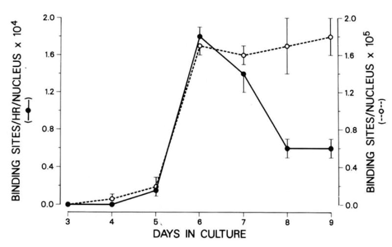
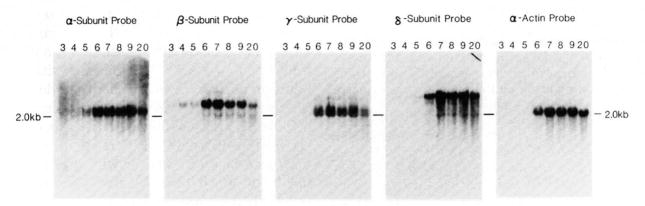
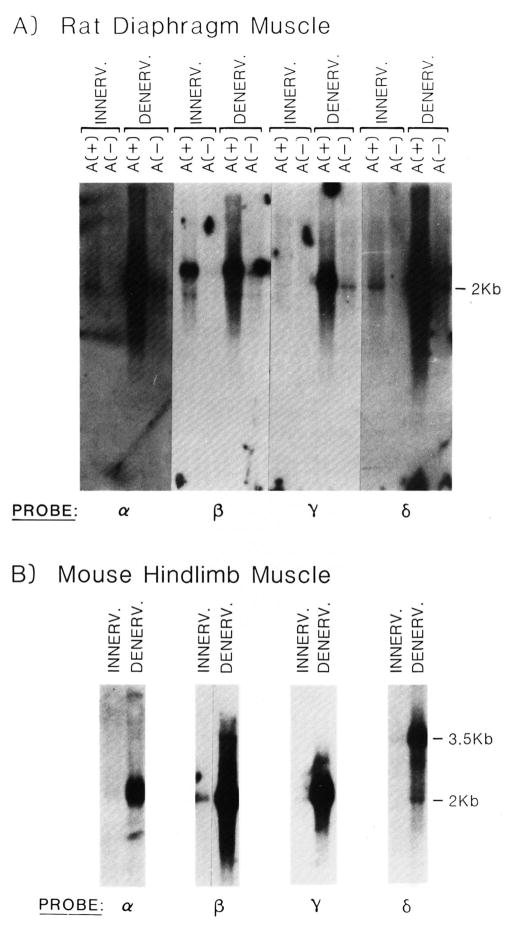
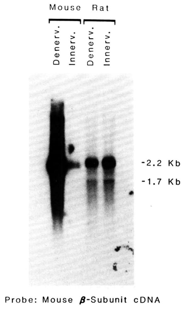
The expression of the muscle nicotinic acetylcholine receptor (AChR) on the cell surface entails a complex biosynthetic pathway, involving the expression and assembly of four subunits. The amount of AChR on the cell surface changes throughout muscle development and upon muscle denervation. We have examined the regulatory role of transcript levels on surface AChR expression by RNA blot analysis. During myogenesis of the fusing mouse muscle cell line C2, which expresses an embryonic type of receptor, changes in the rate of appearance of cell surface AChR have been assayed by 125I-alpha-bungarotoxin binding. The maximal increase in the rate of appearance of cell surface AChR occurs upon cell fusion, closely following the maximal increase in transcript levels for the alpha-, beta-, gamma-, and delta-AChR subunits. AChR alpha-, beta-, gamma-, and delta-subunit gene transcript levels have also been examined in innervated and denervated rat and mouse muscle. Muscle denervation results in an increase of transcripts for all four subunits. However, the amount of beta-subunit transcript in innervated rat skeletal muscle is high relative to the other subunit transcripts, and increases less than the other subunit transcripts upon denervation. Our results indicate that, during myogenesis and upon denervation, the availability of AChR subunit transcripts for translation plays a regulatory role in surface AChR appearance.


Figures

Fig. 1
Fig. 1. The rate of appearance of 125I-α-bungarotoxin binding sites and the total number of binding sites present during the development of C2 cells
The measurement of the rate of appearance of surface acetylcholine receptors and the procedure for 125I-α-bungarotoxin binding were performed as described under “Experimental Procedures.” Each time point was performed on duplicate dishes and data shown are averages of two separate experiments. The error bars indicate the standard deviation between values from separate experiments. Each AChR molecule has two binding sites for α-bungarotoxin. The maximal number of receptors agrees well with the maximal number of receptors reported earlier for C2 cells (Inestrosa et al., 1983). Cells were harvested on day 5 before the addition of fusion medium on that day. Day 6 cultures had been in fusion medium for 24 h.
Fig. 2
Fig. 2. RNA blot analysis of the accumulation of AChR subunit mRNAs during C2 cell myogenesis
Total cellular RNA was extracted from C2 cells and purified as described under “Experimental Procedures.” Ten μg of total RNA from each of the developmental days indicated above each lane (3–9, 20) were run on 1.5% formaldehyde agarose gels, blotted, and probed as described under “Experimental Procedures.” Molecular sizes were determined by comparison to molecular size standards of λ-DNA cut with HindIII, and ϕX174 DNA digested with HaeIII.
Fig. 3
Fig. 3. Quantitation by RNA blot analysis of cytoplasmic RNA from C2 cells before (day 5) and after (day 6) the addition of fusion medium
Cytoplasmic poly(A)+ RNA was prepared from C2 cells at day 5 and day 6 as described under “Experimental Procedures.” RNA blot analysis was performed. The resulting autoradiograms are shown. The amounts of cytoplasmic poly(A)+ RNA loaded from each day are indicated above the autoradiograms of each blot. Two- or four-fold more RNA was loaded for day 5 RNAs relative to day 6 RNAs. The probes used are indicated above the autoradiogram of each blot.
Fig. 4
Fig. 4. Effect of muscle denervation on the levels of mRNA coding for the α-, β-, γ-, and δ-subunits of the acetylcholine receptor
The denervation data shown are from 5-day denervated muscle tissue. Time course experiments indicated that RNA levels for rat muscle were maximal at 4 days postdenervation and maintained throughout a 12-day period (data not shown). A, RNA blot analysis of innervated or 5-day denervated rat diaphragm RNA. Each lane contains approximately 5 μg of either poly(A)+ or -(A) RNA. The circular signal appearing in the poly(A) RNA lane from the denervated tissue probed with the β-subunit probe is an artifact. B, RNA blot analysis of innervated and 5-day denervated mouse hindlimb muscle RNA. Each lane contains approximately 5 μg of poly(A)+ RNA, except those lanes probed with the α-subunit cDNA which contains 4.6 μg of innervated muscle RNA and 3.5 μg of denervated muscle RNA. PROBE refers to one of the four different acetylcholine receptor subunits which were radiolabeled by nick-translation and used to identify their homologous transcript. Specific activities for different probes and the length of time the blot was exposed to x-ray film varied. The blots were exposed for a length of time sufficient to be able to visualize the subunit transcripts from innervated muscle.
Fig. 5
Fig. 5. The effect of denervating the plantar extensor group of muscles of rat or mouse on the level of mRNA coding for the β-subunit of the AChR
5 μg of poly(A)+ RNA isolated from innervated and 5-day denervated plantar extensor muscles of the rat or mouse were applied to the gel. RNA blots were probed with nick-translated 32P-labeled cDNA corresponding to the β-subunit of the AChR. Blots were exposed long enough to enable visualization of innervated muscle transcripts. Specific activities for different probes and the length of time the blot was exposed to x-ray film varied.

References

    1. Arnheim N. Gene ( Amst ) 1979;7:83–96. - PubMed
    1. Aviv H, Leder P. Proc Natl Acad Sci U S A. 1972;69:1408–1412. - PMC - PubMed
    1. Boulter J, Luyten W, Evans K, Mason P, Ballivet M, Goldman D, Stengelin S, Martin G, Heinemann S, Patrick J. J Neurosci. 1985;5:2545–2552. - PMC - PubMed
    1. Boulter J, Evans K, Martin G, Mason P, Stengelin S, Goldman D, Heinemann S, Patrick J. J Neurosci Res. 1986;16:37–49. - PubMed
    1. Buonanno A, Merlie JP. J Biol Chem. 1986;261:11452–11455. - PubMed