. 2013 Dec 20;342(6165):1521-4.
doi: 10.1126/science.1244142.

Serial femtosecond crystallography of G protein-coupled receptors

Affiliations

Serial femtosecond crystallography of G protein-coupled receptors

Wei Liu et al. Science. .

Abstract

X-ray crystallography of G protein-coupled receptors and other membrane proteins is hampered by difficulties associated with growing sufficiently large crystals that withstand radiation damage and yield high-resolution data at synchrotron sources. We used an x-ray free-electron laser (XFEL) with individual 50-femtosecond-duration x-ray pulses to minimize radiation damage and obtained a high-resolution room-temperature structure of a human serotonin receptor using sub-10-micrometer microcrystals grown in a membrane mimetic matrix known as lipidic cubic phase. Compared with the structure solved by using traditional microcrystallography from cryo-cooled crystals of about two orders of magnitude larger volume, the room-temperature XFEL structure displays a distinct distribution of thermal motions and conformations of residues that likely more accurately represent the receptor structure and dynamics in a cellular environment.


Figures

Fig. 1
Fig. 1. Experimental setup for SFX data collection using an LCP injector
5-HT2B receptor microcrystals (first zoom level) dispersed in LCP (second zoom level) are injected as a continuous column of 20–50 µm diameter inside a vacuum chamber and intersected with 1.5 µm diameter pulsed XFEL beam focused by Kirkpatrick-Baez (K-B) mirrors. Single pulse diffraction patterns are collected at 120 Hz using a CSPAD detector.
Fig. 2
Fig. 2. Comparison between 5-HT2B-XFEL (light red) and 5-HT2B-SYN (cyan) structures
Central image represents a backbone overlay of the two structures. Dashed lines correspond to membrane boundaries defined by the Orientation of Proteins in Membrane database (http://opm.pharm.umich.edu) (28). (A) Electron density for the Glu212 side chain is missing in 5-HT2B-SYN and fully resolved in 5-HT2B-XFEL. (B) A salt bridge between Glu319 and Lys247 links intracellular parts of helices V and VI in the 5-HT2B-XFEL structure. In the 5-HT2B-SYN structure Lys247 makes a hydrogen bond with Tyr1105 from the BRIL fusion protein. (C) Extracellular tip of helix II forms a regular helix in 5-HT2B-XFEL with Thr114 making a stabilizing hydrogen bond with the backbone carbonyl, while in 5-HT2B-SYN a water stabilized kink is introduced at this position. (D) Tyr87 forms a hydrogen bond with Asn90 in 5-HT2B-XFEL; this hydrogen bond is broken and Tyr87 adopts a different rotamer conformation in 5-HT2B-SYN structure. 2mFobs-Fcalc maps (contoured at 1 σ level) are shown only around described residues.
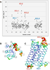
Fig. 3
Fig. 3. Differences in B-factors between 5-HT2B-XFEL and 5-HT2B-SYN structures
(A) B-factors difference (BXFEL-BSYN) for Cα atoms plotted vs. residue number. (B) View of the 5-HT2B-XFEL structure from the extracellular side and (C) in the lateral to membrane orientation. Structure in B and C is shown in putty representation and colored in rainbow colors by the Cα B-factors (range 60 – 170 Å2). Loops for which B-factors increased above average are labeled in red and those that have about an average increase are labeled in blue in (A) and (C). Helices are labeled in (B).

Comment in

  • Making protein crystals fly.
    Doerr A. Doerr A. Nat Methods. 2014 Apr;11(4):366-7. doi: 10.1038/nmeth.2913. Nat Methods. 2014. PMID: 24818225 No abstract available.
  • Serial femtosecond crystallography datasets from G protein-coupled receptors.
    White TA, Barty A, Liu W, Ishchenko A, Zhang H, Gati C, Zatsepin NA, Basu S, Oberthür D, Metz M, Beyerlein KR, Yoon CH, Yefanov OM, James D, Wang D, Messerschmidt M, Koglin JE, Boutet S, Weierstall U, Cherezov V. White TA, et al. Sci Data. 2016 Aug 1;3:160057. doi: 10.1038/sdata.2016.57. Sci Data. 2016. PMID: 27479354 Free PMC article.

References

    1. Cherezov V, et al. High-resolution crystal structure of an engineered human beta2-adrenergic G protein-coupled receptor. Science. 2007;318:1258–1265. - PMC - PubMed
    1. Rasmussen SG, et al. Crystal structure of the beta(2) adrenergic receptor-Gs protein complex. Nature. 2011;469:175–180. - PMC - PubMed
    1. Katritch V, Cherezov V, Stevens RC. Structure-function of the G protein-coupled receptor superfamily. Ann. Rev. Pharmacol Toxicol. 2013;53:531–556. - PMC - PubMed
    1. Venkatakrishnan AJ, et al. Molecular signatures of G-protein-coupled receptors. Nature. 2013;494:185–194. - PubMed
    1. Audet M, Bouvier M. Restructuring G-protein- coupled receptor activation. Cell. 2012;151:14–23. - PubMed

Publication types

MeSH terms

Substances

Associated data