

Metal shadowing for electron microscopy
- PMID: 24357360
- PMCID: PMC7120032
- DOI: 10.1007/978-1-62703-776-1_5
Metal shadowing for electron microscopy
Abstract
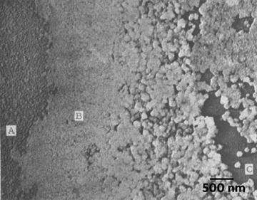
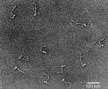
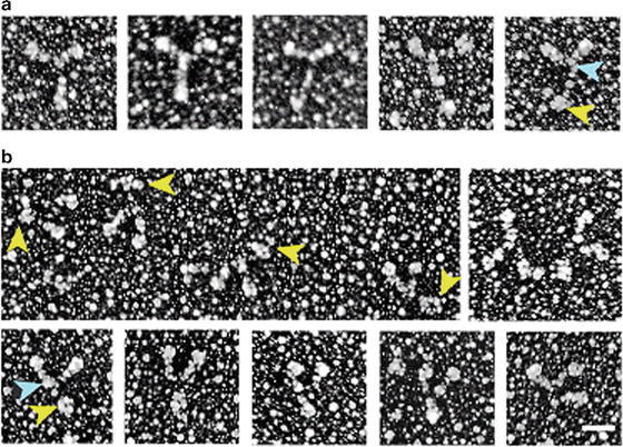
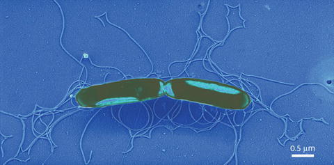
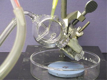
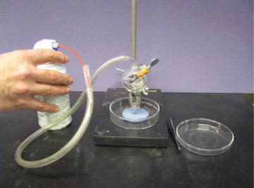
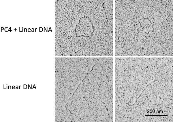
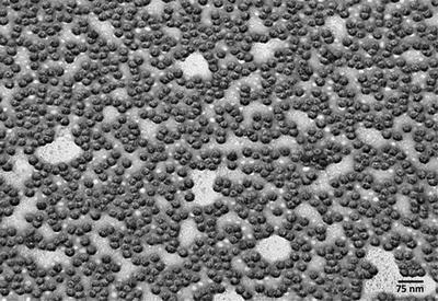
Metal shadowing of bacteria, viruses, isolated molecules, and macromolecular assemblies is another high-resolution method for observing the ultrastructure of biological specimens. The actual procedure for producing a metal shadow is relatively simple; a heavy metal is evaporated from a source at an oblique angle to the specimen. The metal atoms pile up on the surfaces that face the source, but the surfaces away from the source are shielded and receive little metal deposit, creating a "shadow." However, the process of producing biological specimens that are suitable for metal shadowing can be very complex. There are a whole host of specimen preparation techniques that can precede metal shadowing, and all provide superior preservation in comparison to air drying, a required step in negative staining procedures. The physical forces present during air drying (i.e., surface tension of the water-air interface) will literally crush most biological specimens as they dry. In this chapter I explain the development of and procedures for the production of biological specimens from macromolecular assemblies (e.g., DNA and RNA), purified isolated molecules (e.g., proteins), and isolated viruses and bacteria preparations suitable for metal shadowing. A variation on this basic technique is to rotate the specimen during the metal deposition to produce a high-resolution three-dimensional rendering of the specimen.
Figures
Similar articles
-
Sputter shadowing.J Ultrastruct Res. 1984 May;87(2):97-105. doi: 10.1016/s0022-5320(84)80069-6. J Ultrastruct Res. 1984. PMID: 6544872
-
Image interpretation of metal shadowed specimens with the aid of Monte Carlo simulation of the shadowing process.Scanning Microsc Suppl. 1988;2:379-86. Scanning Microsc Suppl. 1988. PMID: 3244974
-
Preparation of viral samples within biocontainment for ultrastructural analysis: Utilization of an innovative processing capsule for negative staining.J Virol Methods. 2016 Dec;238:70-76. doi: 10.1016/j.jviromet.2016.10.005. Epub 2016 Oct 14. J Virol Methods. 2016. PMID: 27751950
-
Determining the structure of biological macromolecules by transmission electron microscopy, single particle analysis and 3D reconstruction.Prog Biophys Mol Biol. 2001;75(3):121-64. doi: 10.1016/s0079-6107(01)00004-9. Prog Biophys Mol Biol. 2001. PMID: 11376797 Review.
-
The use of trehalose in the preparation of specimens for molecular electron microscopy.Micron. 2011 Dec;42(8):762-72. doi: 10.1016/j.micron.2011.06.005. Epub 2011 Jun 25. Micron. 2011. PMID: 21752659 Free PMC article. Review.
Cited by
-
Fine structure of the vaccinia virion determined by controlled degradation and immunolocalization.Virology. 2015 Jan 15;475:204-18. doi: 10.1016/j.virol.2014.11.020. Epub 2014 Dec 8. Virology. 2015. PMID: 25486587 Free PMC article.
References
-
- Marton L. La microscopie electronique des onjectes biologiques. Bull Acad Belg Cl Sci. 1934;20:439–466.
-
- Schaefer FJ, Harker DJ. Surface replicas for use in the electron microscope. J Appl Phys. 1942;13:427–433. doi: 10.1063/1.1714890. - DOI
-
- Sharp DG, Lanni F, Beard JW. The egg white inhibitor of influenza virus hemagglutination. J Biol Chem. 1950;185:681–688. - PubMed
-
- Heinmets F. Modification of silica replica technique for study of biological membranes and application of rotary condensation in electron microscopy. J Appl Physiol. 1949;20:384–388. doi: 10.1063/1.1698372. - DOI
MeSH terms
Substances
LinkOut - more resources
Full Text Sources
Other Literature Sources
