. 2014 Jan 7;33(1):76-92.
doi: 10.1002/embj.201386124. Epub 2013 Dec 15.

The mechanism of translation initiation on Type 1 picornavirus IRESs

Affiliations

The mechanism of translation initiation on Type 1 picornavirus IRESs

Trevor R Sweeney et al. EMBO J. .

Abstract

Picornavirus Type 1 IRESs comprise five principal domains (dII-dVI). Whereas dV binds eIF4G, a conserved AUG in dVI was suggested to stimulate attachment of 43S ribosomal preinitiation complexes, which then scan to the initiation codon. Initiation on Type 1 IRESs also requires IRES trans-acting factors (ITAFs), and several candidates have been proposed. Here, we report the in vitro reconstitution of initiation on three Type 1 IRESs: poliovirus (PV), enterovirus 71 (EV71), and bovine enterovirus (BEV). All of them require eIF2, eIF3, eIF4A, eIF4G, eIF4B, eIF1A, and a single ITAF, poly(C) binding protein 2 (PCBP2). In each instance, initiation starts with binding of eIF4G/eIF4A. Subsequent recruitment of 43S complexes strictly requires direct interaction of their eIF3 constituent with eIF4G. The following events can differ between IRESs, depending on the stability of dVI. If it is unstructured (BEV), all ribosomes scan through dVI to the initiation codon, requiring eIF1 to bypass its AUG. If it is structured (PV, EV71), most initiation events occur without inspection of dVI, implying that its AUG does not determine ribosomal attachment.


Figures

Figure 1
Figure 1
Factor requirements for initiation at the 3’-border of the PV IRES. A  Model of the PV1M IRES (Genbank V01149). Inset panel shows mutations (red letters) and the structure of dVI in AUG586-good mRNA. B–E  Toeprinting analysis of 48S complex formation at AUG586 on (B, C, E) AUG586-good mRNA, (D) AUG586-good mRNA containing the C299A substitution. F  AUG586-good mRNA containing mutations in domain V. G  Binding of eIF4G to AUG586-good mRNA containing mutations in domain V as indicated in (F), analyzed by HRC using the eIF4Gm(T829C) mutant. Cleavage sites are indicated on the right. H, I  Toeprinting analysis of 48S complex formation at AUG586 on (H) AUG586-good mRNA containing mutations in domain V and (I) AUG586-good mRNA containing mutations in dVI shown in the left panel. Data information: For (B–E, H, I), 48S complexes were formed at 100 mM K+. Reaction mixtures contained 40S subunits, Met-tRNAiMet and indicated sets of eIFs and ITAFs. Toeprints caused by binding of GARS, La or PCBP2 and by 48S complexes assembled at AUG586 are shown on the right. The division between sequence lanes and lanes 1–10 in (H) indicates that these two sets of lanes were derived from the same gel, exposed for different lengths of time.
Figure 2
Figure 2
48S complex formation at the PV initiation codon AUG743. A–F  Toeprinting analysis of 48S complex formation on (A, left panel; B–E) PV wt mRNA, (A, right panel) AUG586 (3′+5′ stabilized) mRNA (Fig 1I), and (F) AUG586/611/683/743 mRNA (F, upper panel). 48S complexes were formed at indicated K+ concentrations. Reaction mixtures contained 40S subunits, Met-tRNAiMet and indicated sets of eIFs and ITAFs. Toeprints caused by 48S complexes assembled at AUG586, AUG611, AUG683 and AUG743 are shown on the right. G  Analysis of 80S ribosomal complexes formed on AUG586/611/683/743 mRNA in RRL in the presence of cycloheximide. After assembly, ribosomal complexes were separated by SDG centrifugation, and fractions corresponding to 80S ribosomes were assayed by toe-printing (lanes 1–2) or RelE cleavage (lanes 3–4). Positions of toe-prints and RelE cleavages corresponding to ribosomal complexes formed at AUG586, AUG611, AUG683 and AUG743 are indicated. Data information: The divisions between sequence lanes and toe-printing lanes in (A, left panel), (C), (D) and (G) indicate that the two sets of lanes shown in each panel were derived from the same gel, exposed for different lengths of time.
Figure 3
Figure 3
48S complex formation on EV71 and BEV IRESs. A  Models of dVI in wt PV, EV71 and BEV IRESs. B–G  Toe-printing analysis of 48S complex formation on (B) wt EV71 mRNA, (C) wt BEV mRNA, (D) AUG668→CUA BEV mRNA (left panel), (E) EV71 mRNAs containing destabilizing mutations in dVI (upper panel), (F) EV71 mRNA containing the additional AUG604 (upper panel), and (G) EV71 mRNAs containing additional AUG triplets at positions 649, 661, 674 or 699. 48S complexes were formed at 100 mM K+. Reaction mixtures contained 40S subunits, Met-tRNAiMet and indicated sets of eIFs and ITAFs. Asterisks show toe-prints caused by 48S complexes assembled on near-cognate initiation codons. Toeprints caused by 48S complexes assembled on AUG triplets are indicated by arrows. The division between sequence lanes and lanes 1–5 in (B) indicates that these two sets of lanes were derived from the same gel, exposed for different lengths of time.
Figure 4
Figure 4
Influence of mutations in domain VI on 48S complex formation on PV and EV71 IRESs. A, B  Mutations (red) introduced into dVI of (A) PV and (B) EV71 IRESs. C–F  Translation of mRNAs containing mutations in dVI of PV, EV71 (A and B) and BEV (Fig 3D) IRESs in RRL, HeLa cell extract, and RRL supplemented with HeLa cell extract (30%, v/v), as indicated. Translation products were quantified relative to those of wt mRNAs, which were defined as 100%. G–I  Toe-printing analysis of 48S complex formation on EV71 mRNAs containing mutations in dVI (B). 48S complexes were formed at 100 mM K+. Reaction mixtures contained 40S subunits, Met-tRNAiMet and indicated sets of eIFs and PCBP2. Asterisks show toe-prints caused by 48S complexes assembled on near-cognate initiation codons. Toeprints caused by 48S complexes assembled on AUG triplets are indicated by arrows.
Figure 5
Figure 5
Interaction of PCBP2 with PV and EV71 IRESs.
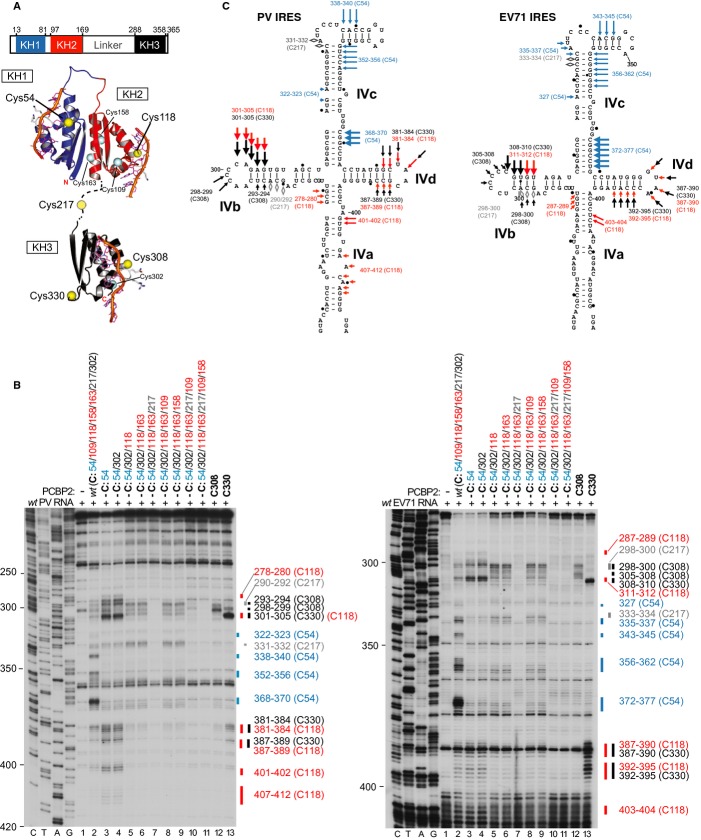
  1.   Schematic representation of PCBP2 showing the positions of the three KH domains (upper panel). Ribbon diagrams of PCBP2 KH1-KH2 and KH3 domains (PDB: 2JZX and 2P2R) with the linker between them represented by a dashed line (lower panel). Spheres indicate native (C54, C109, C118, C158, C163, C207, C302) and introduced (C308, C330) cysteines. Cysteines that induced cleavage are colored yellow.

  2.   Primer extension analysis after HRC of PV (left) and EV71 (right) IRESs from Fe(II)-tethered PCBP2.

  3.   Models of the secondary structures of apical subdomains IVa-IVd of PV and EV71 IRESs, marked to show sites of HRC from PCBP2. Arrow size is proportional to cleavage intensity. Sites of HRC (B, C) are colored to match domains in (A).

Figure 6
Figure 6
Direct interaction of eIF4G with eIF3 is required for recruitment of 43S complexes to the PV IRES. A  Schematic representation of full-length and truncated eIF4G1, showing sites of factor binding and of cleavage by 2Apro. B  Recruitment of eIF4A to the PV IRES by eIF4G mutants, analyzed by HRC using the eIF4A(S42C) mutant. Cleavage sites are indicated on the right. C–E  Toeprint analysis of 48S complex formation on (C) AUG586-good PV, (D) wt PV and (E) EMCV mRNAs, in reaction mixtures that contained 40S subunits, Met-tRNAiMet, eIFs, ITAFs and eIF4F, eIF4G653–1599, eIF4G736–1115, eIF4G736–1008 or eIF4G736–988, as indicated. 48S complexes were formed at indicated K+ concentrations. Toeprints caused by 48S complexes assembled at EMCV AUG826 and AUG834, PV AUG586 and PV AUG743 are indicated on the right.
Figure 7
Figure 7
Position of eIF3 on Type 1 IRESs assayed by directed hydroxyl radical cleavage. A–D  Primer extension analysis of HRC of (A) CSFV, (B, D) PV and (C) EV71 IRESs from Fe(II)-tethered native eIF3 in the presence of eIF4G736–1115, eIF4G736–1008, eIF4G736–988 and eIF4A, as indicated. Cleavage sites are shown on the right and are mapped onto structural models of CSFV, PV and EV71 IRESs (A–C, lower panels). The division between sequence lanes and lanes 1–5 in (A) indicates that these two sets of lanes were derived from the same gel, exposed for different lengths of time.
Figure 8
Figure 8
Stimulation by eIF3 of eIF4G's interaction with Type 1 IRESs. A–C  Primer extension analysis of directed hydroxyl radical cleavage of (A) EV71, (B) PV and (C) EMCV IRESs from Fe(II)-tethered eIF4Gm(T829C) in the presence of eIF3 and/or eIF4A as indicated. Cleavage sites are shown on the right.

References

    1. Abaeva IS, Marintchev A, Pisareva VP, Hellen CU, Pestova TV. Bypassing of stems versus linear base-by-base inspection of mammalian mRNAs during ribosomal scanning. EMBO J. 2011;30:115–129. - PMC - PubMed
    1. Anderson EC, Hunt SL, Jackson RJ. Internal initiation of translation from the human rhinovirus-2 internal ribosome entry site requires the binding of Unr to two distinct sites on the 5′ untranslated region. J Gen Virol. 2007;88:3043–3052. - PubMed
    1. Andreev DE, Fernandez-Miragall O, Ramajo J, Dmitriev SE, Terenin IM, Martinez-Salas E, Shatsky IN. Differential factor requirement to assemble translation initiation complexes at the alternative start codons of foot-and-mouth disease virus RNA. RNA. 2007;13:1366–1374. - PMC - PubMed
    1. Andreev D, Hauryliuk V, Terenin I, Dmitriev S, Ehrenberg M, Shatsky I. The bacterial toxin RelE induces specific mRNA cleavage in the A site of the eukaryote ribosome. RNA. 2008;14:233–239. - PMC - PubMed
    1. Andreev DE, Hirnet J, Terenin IM, Dmitriev SE, Niepmann M, Shatsky IN. Glycyl-tRNA synthetase specifically binds to the poliovirus IRES to activate translation initiation. Nucleic Acids Res. 2012;40:5602–5614. - PMC - PubMed

Publication types

MeSH terms