

Type 1 metabotropic glutamate receptors (mGlu1) trigger the gating of GluD2 delta glutamate receptors
- PMID: 24357660
- PMCID: PMC4303454
- DOI: 10.1002/embr.201337371
Type 1 metabotropic glutamate receptors (mGlu1) trigger the gating of GluD2 delta glutamate receptors
Abstract
The orphan GluD2 receptor belongs to the ionotropic glutamate receptor family but does not bind glutamate. Ligand-gated GluD2 currents have never been evidenced, and whether GluD2 operates as an ion channel has been a long-standing question. Here, we show that GluD2 gating is triggered by type 1 metabotropic glutamate receptors, both in a heterologous expression system and in Purkinje cells. Thus, GluD2 is not only an adhesion molecule at synapses but also works as a channel. This gating mechanism reveals new properties of glutamate receptors that emerge from their interaction and opens unexpected perspectives regarding synaptic transmission and plasticity.
Figures
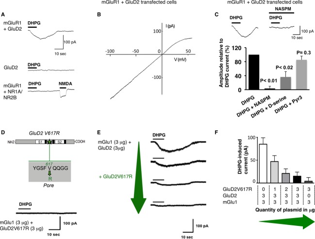
DHPG induced current at −60 mV in cells expressing mGlu1, GluD2 or NR1-NR2B subunits, alone or in combination.
The I–V relationship of the DHPG current in a cell expressing mGlu1 and GluD2.
NASPM, D-serine but not Pyr3 inhibit the DHPG current in cells transfected with GluD2 and mGlu1.
The GluD2V617R dominant-negative construct (top, S1, S2: LBD). Lack of DHPG current in cells expressing mGlu1 and GluD2V617R (bottom).
GluD2V617R reduces the DHPG current. Representative traces from cells transfected with mGlu1 and GluD2 together with increasing amounts of GluD2V617R plasmid (green arrow).
Averaged amplitude of DHPG currents (± s.e.m.) as a function of the quantity of plasmids co-transfected as indicated below.
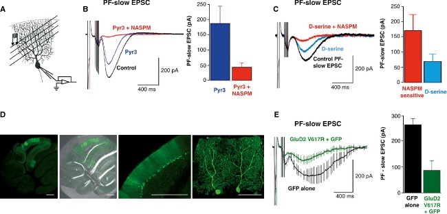
A Experimental arrangement for the mGlu1 synaptic activation.
B,C mGlu1-slow ESPCs are inhibited by Pyr3, NASPM (B) and D-serine (C). Representative sweeps. Histograms show the PF-slow EPSC averaged peak amplitudes.
D A slice from WT cerebellum infected with Sinbis carrying GFP and GluD2V617R. Scale bars: 500 μm (three left images), 100 μm (right).
E Averaged PF-slow EPSC from V617RGluD2 (green) or GFP (black) expressing cells. Histograms: Corresponding PF-slow EPSCs averaged peak amplitudes.
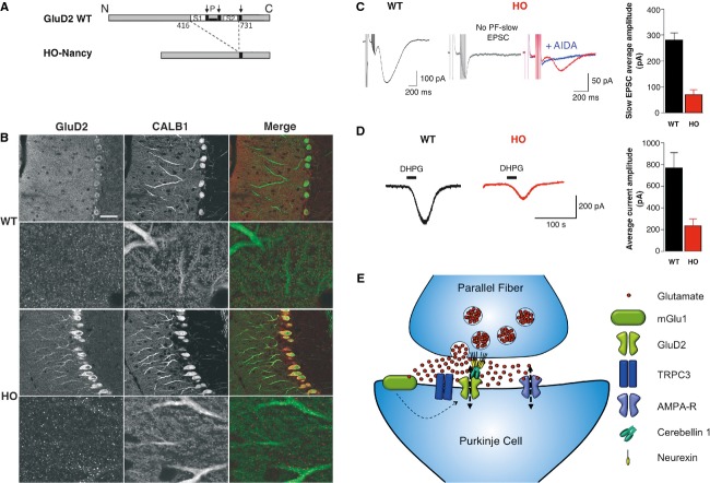
A WT and HO-Nancy schematic proteins. Amino-acid numbers flanking the deletion are indicated. Arrows: transmembrane domains. P: channel pore.
B Confocal images of calbindin and GluD2 immunolabelings in WT (top) and HO-Nancy (bottom) cerebella. Scale bar in top left image represents 45 μm.
C,D Representative PF-slow ESPCs (C) and DHPG currents (D) from WT (left) and HO-Nancy (right) Purkinje cells. Histograms: averaged peak amplitude of PF-slow ESPCs (C) or DHPG currents (D) from all the cells (± s.e.m.).
E Schematic representation of PF synapses. GluD2s are not directly activated by glutamate (red spots) but require mGlu1 activation and thus presynaptic PF bursts.
References
-
- Kohda K, Wang Y, Yuzaki M. Mutation of a glutamate receptor motif reveals its role in gating and delta2 receptor channel properties. Nat Neurosci. 2000;3:315–322. - PubMed
-
- Lomeli H, Sprengel R, Laurie DJ, Kohr G, Herb A, Seeburg PH, Wisden W. The rat delta-1 and delta-2 subunits extend the excitatory amino acid receptor family. FEBS Lett. 1993;315:318–322. - PubMed
-
- Kashiwabuchi N, Ikeda K, Araki K, Hirano T, Shibuki K, Takayama C, Inoue Y, Kutsuwada T, Takeshi Yagi T, Kang Y, Aizawa S, Mishina M. Impairement of motor coordination, Purkinje cell synapses formation, and cerebellar long-term depression in GluR-delta-2 mutant mice. Cell. 1995;81:245–252. - PubMed
-
- Filali M, Lalonde R, Bensoula AN, Guastavino JM, Lestienne F. Spontaneous alternation, motor activity, and spatial learning in hot-foot mutant mice. J Comp Physiol A. 1996;178:101–104. - PubMed
Publication types
MeSH terms
Substances
LinkOut - more resources
Full Text Sources
Other Literature Sources
Molecular Biology Databases
