. 2014 Jun;25(6):1211-25.
doi: 10.1681/ASN.2013080831. Epub 2013 Dec 19.

Rapid and efficient differentiation of human pluripotent stem cells into intermediate mesoderm that forms tubules expressing kidney proximal tubular markers

Affiliations

Rapid and efficient differentiation of human pluripotent stem cells into intermediate mesoderm that forms tubules expressing kidney proximal tubular markers

Albert Q Lam et al. J Am Soc Nephrol. 2014 Jun.

Abstract

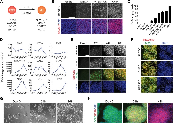
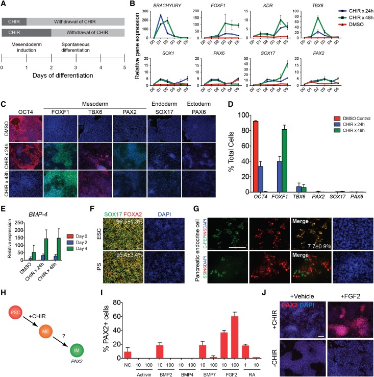
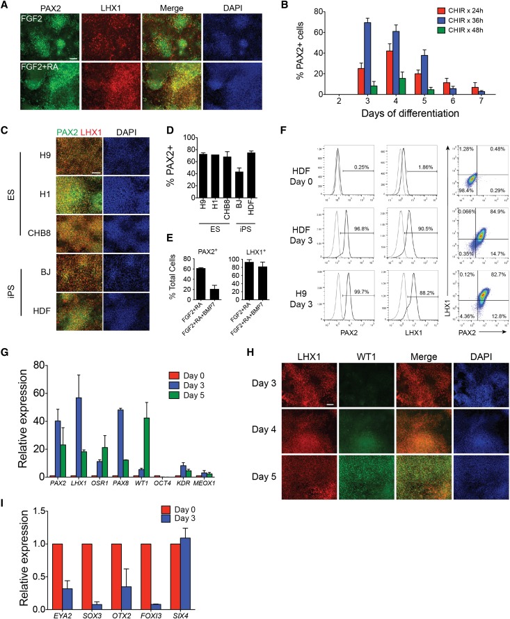
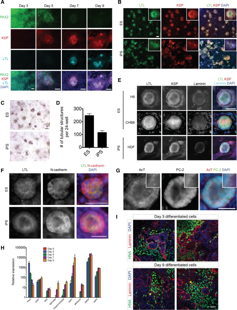
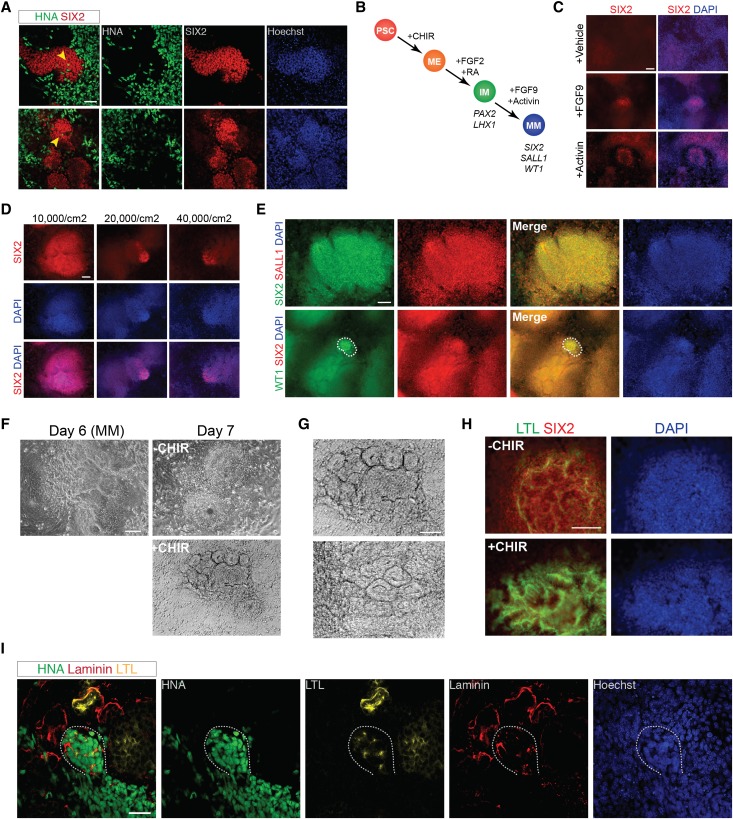
Human pluripotent stem cells (hPSCs) can generate a diversity of cell types, but few methods have been developed to derive cells of the kidney lineage. Here, we report a highly efficient system for differentiating human embryonic stem cells and induced pluripotent stem cells (referred to collectively as hPSCs) into cells expressing markers of the intermediate mesoderm (IM) that subsequently form tubule-like structures. Treatment of hPSCs with the glycogen synthase kinase-3β inhibitor CHIR99021 induced BRACHYURY(+)MIXL1(+) mesendoderm differentiation with nearly 100% efficiency. In the absence of additional exogenous factors, CHIR99021-induced mesendodermal cells preferentially differentiated into cells expressing markers of lateral plate mesoderm with minimal IM differentiation. However, the sequential treatment of hPSCs with CHIR99021 followed by fibroblast growth factor-2 and retinoic acid generated PAX2(+)LHX1(+) cells with 70%-80% efficiency after 3 days of differentiation. Upon growth factor withdrawal, these PAX2(+)LHX1(+) cells gave rise to apically ciliated tubular structures that coexpressed the proximal tubule markers Lotus tetragonolobus lectin, N-cadherin, and kidney-specific protein and partially integrated into embryonic kidney explant cultures. With the addition of FGF9 and activin, PAX2(+)LHX1(+) cells specifically differentiated into cells expressing SIX2, SALL1, and WT1, markers of cap mesenchyme nephron progenitor cells. Our findings demonstrate the effective role of fibroblast growth factor signaling in inducing IM differentiation in hPSCs and establish the most rapid and efficient system whereby hPSCs can be differentiated into cells with features characteristic of kidney lineage cells.


Figures

Figure 1.
Figure 1.
CHIR99021 efficiently induces human pluripotent stem cells to differentiate into mesendoderm-like cells. (A) Diagram of differentiation of human PSCs into mesendoderm using CHIR. (B) hPSCs treated with DMSO (vehicle), Wnt3a 500 ng/ml, Wnt3a 500 ng/ml+activin 50 ng/ml, or CHIR 5 μM were immunostained for BRACHYURY after 24 hours of differentiation. (C) Quantification of cells with positive immunofluorescence staining for BRACHYURY after treatment with vehicle, Wnt3a at increasing doses, Wnt3a at increasing doses with activin, or CHIR for 24 hours. Data represent mean±SEM (n=3). (D) Time course of gene expression in hPSCs treated with CHIR. Quantitative RT-PCR of genes expressed in mesendoderm and in pluripotent stem cells. Data represent mean±SEM (n>3). (E) Immunofluorescence staining for BRACHYURY and MIXL1 in CHIR-treated hPSCs over the first 48 hours of differentiation. (F) Immunofluorescence staining for BRACHYURY and MIXL1 after 24 hours of differentiation in hESC and hiPSC lines treated with CHIR. (G) Phase contrast images of CHIR-treated human PSCs over 36 hours showing epithelial to mesenchymal transition. (H) Immunofluorescence staining for E-cadherin and N-cadherin in CHIR-treated human PSCs. Scale bars, 100 μm. BRACHY, BRACHYURY; CHIR, CHIR99021; ME, mesendoderm; PSC, pluripotent stem cell. BJ, iPS cells derived from human foreskin fibroblasts; DAPI, 4′,6-diamidino-2-phenylindole; HDF, iPS cells derived from adult human dermal fibroblasts.
Figure 2.
Figure 2.
Timed addition of exogenous factors modulates cell fate of CHIR-induced hPSCs. (A) Diagram depicting time course of differentiation. (B) Time course of gene expression in human PSCs treated with CHIR for 24 hours, CHIR for 48 hours, or DMSO (vehicle). Data shown represent mean±SEM (n=4). Representative immunostaining (C) and quantification (D) of markers of pluripotency, mesoderm, definitive endoderm, and ectoderm in human PSCs treated with DMSO (vehicle), CHIR for 24 hours, or CHIR for 48 hours, day 4 of differentiation. Data shown in graph represent mean±SEM (n>5). (E) Expression of BMP-4 by quantitative RT-PCR in hPSCs treated with CHIR for 24 hours, CHIR for 48 hours, or DMSO. Data shown represent mean±SEM (n=3). (F) Representative immunostaining for SOX17 and FOXA2 in hESCs and hiPSCs treated with CHIR for 24 hours followed by activin A 100 ng/ml for 3 days. Numbers represent the mean percentage±SEM of SOX17+ cells from at least two independent experiments. (G) Cells at the definitive endoderm stage were differentiated using a three-step protocol into hormone-expressing pancreatic endocrine cells producing insulin, proinsulin C-peptide, and somatostatin. Number represents the mean percentage±SEM of insulin+C-peptide+ cells from at least two independent experiments. (H) Diagram of directed differentiation of hPSCs into intermediate mesoderm. (I) Quantification of cells with positive immunostaining for PAX2. Data shown in the graph represent mean±SEM (n=2). (J) Immunostaining for PAX2 in hPSCs treated with or without CHIR for 24 hours followed by FGF2 100 ng/ml for 3 days, day 4. Scale bars, 100 μm. DAPI, 4′,6-diamidino-2-phenylindole.
Figure 3.
Figure 3.
FGF2 and RA induce PAX2+LHX1+ intermediate mesoderm cells. (A) Immunostaining for PAX2 and LHX1 in hPSCs cells treated with CHIR for 24 hours followed by FGF2 or FGF2+RA for 3 days, day 4 of differentiation. (B) Quantification of PAX2+ cells on days 2–7 of differentiation in hPSCs treated with CHIR for 24, 36, or 48 hours followed by FGF2+RA. Data shown represent mean±SEM (n>4). (C) Immunostaining for PAX2 and LHX1 and (D) quantification of PAX2+ cells in three hESC lines and two hiPSC lines treated with CHIR for 36 hours followed by FGF2+RA (ChFR), day 3. Data shown represent mean±SEM (n=3). (E) Quantification of PAX2+ or LHX1+ cells in hPSCs treated with CHIR for 36 hours followed by FGF2+RA or FGF2+RA+BMP7, day 3. Data shown represent mean±SEM (n=3). (F) Quantification of PAX2+ and LHX1+ cells by flow cytometry. (G) Quantitative RT-PCR of intermediate mesoderm genes in hPSCs treated with ChFR. Data shown represent mean±SEM (n=3). (H) Immunostaining for LHX1 and WT1 in hPSCs treated with ChFR. (I) Quantitative RT-PCR of non-IM genes in hPSCs treated with ChFR. Data shown represent mean±SEM (n=2). Scale bar, 100 μm. DAPI, 4′,6-diamidino-2-phenylindole.
Figure 4.
Figure 4.
PAX2+LHX1+ intermediate mesoderm cells form polar tubular structures expressing polycystin-2+ cilia and kidney proximal tubular markers. (A) Immunostaining time course from days 3 to 9 for PAX2, KSP, and LTL in hPSCs treated with FGF2+RA (ChFR) for 3 days, then cultured in media without additional growth factors for an additional 6 days. Scale bar, 50 μm. (B) Immunostaining for KSP and LTL in tubular structures formed by PAX2+LHX1+ IM cells, day 9. Inset shows tubular structure at higher magnification. Scale bar, 50 μm. (C) Brightfield stereomicroscope imaging of tubular structures, day 9. Scale bar, 200 μm. (D) Quantification of tubular structures formed from PAX2+LHX1+ IM cells, day 9. Data shown represent mean±SEM (n=4 for one hESC and n=4 for one hiPSC line). (E) Immunostaining for KSP, LTL, and laminin in tubular structures derived from two hESC lines and one iPSC line, day 9. Scale bar, 50 μm. (F) Immunostaining for LTL and N-cadherin in tubular structures derived from one hESC and one iPSC line, day 9. Scale bar, 50 μm. (G) Immunostaining for acetylated α-tubulin and polycystin-2 in tubular structures, day 9. Inset shows higher magnification of cilia localized to the apical surface. Scale bar, 50 μm. (H) Quantitative RT-PCR of genes associated with kidney development and mature kidney epithelial cells. Data shown represent mean±SEM (n=2). (I) Whole-mount immunohistochemistry for anti–human nuclear antigen (HNA) and laminin in chimeric kidney explant cultures. Dissociated hPSC-derived IM cells on day 3 (n=10) and day 9 (n=3) of differentiation were mixed with dissociated E12.5 mouse embryonic kidneys in single cell suspension, reaggregated by centrifugation, and cultured for 3 days in kidney explant culture. Arrowhead, laminin-bounded structures containing human and mouse cells. Scale bar, 50 μm. AQP1, aquaporin-1; AQP2, aquaporin-2; DAPI, 4′,6-diamidino-2-phenylindole; HNA, human nuclear antigen; UMOD, uromodulin.
Figure 5.
Figure 5.
FGF-9 and activin differentiate PAX2+LHX1+ cells into cells expressing markers of CM. (A) Whole-mount immunohistochemistry for anti–human nuclear antigen (HNA) and SIX2 in chimeric kidney explant cultures. Dissociated hESC-derived IM cells on day 9 (n=3) of differentiation were mixed with dissociated E12.5 mouse embryonic kidneys in single cell suspension, reaggregated by centrifugation, and cultured for 5 days in kidney explant culture. Arrowhead, HNA+ cells present within clusters of mouse Six2+ cells. Scale bar, 50 μm. (B) Diagram showing the stepwise differentiation of hESCs into metanephric CM. (C) Immunostaining for SIX2 in hESCs treated with FGF2+RA (ChFR) for 3 days then either 100 ng/ml FGF-9, 10 ng/ml activin, or vehicle for 3 days, day 6. Scale bar, 100 μm. (D) Immunostaining for SIX2 in hESCs plated at different densities and treated with ChFR for 3 days then 100 ng/ml FGF-9+10 ng/ml activin for 3 days, day 6. Scale bar, 100 μm. (E) Immunostaining for SIX2, SALL1, and WT1 in hESCs treated with ChFR for 3 days then 100 ng/ml FGF-9+10 ng/ml activin for 3 days, day 6. Dashed line encompasses the population of cells that stain positive for WT1. Scale bar, 100 μm. (F) Brightfield microscopy of hESCs treated with ChFR for 3 days, 100 ng/ml FGF-9+10 ng/ml activin for 3 days, then either 5 μM CHIR (+CHIR) or FGF-9+activin (−CHIR) for 24 hours. Scale bar, 100 μm. (G) Higher magnification of tubular epithelial-like structures seen in SIX2+ cells treated on day 6 with 5 μM CHIR for 24 hours, day 7. Scale bar, 100 μm. (H) Immunostaining (day 8) for SIX2 and LTL in hESCs treated with ChFR for 3 days, 100 ng/ml FGF-9+10 ng/ml activin for 3 days, then either 5 μM CHIR (+CHIR) or FGF9+activin (−CHIR) for 24 hours. Scale bar, 100 μm. (I) Whole-mount immunohistochemistry for anti–human nuclear antigen (HNA), laminin, and LTL in chimeric kidney explant cultures. Dissociated hESC-derived SIX2+ cells on day 6 (n=10) of differentiation were mixed with dissociated E12.5 mouse embryonic kidneys in single cell suspension, reaggregated by centrifugation, and cultured for 3 days in kidney explant culture. Dashed line encompasses an organizing cluster of HNA+ cells, which express laminin and LTL. Scale bar, 50 μm. DAPI, 4′,6-diamidino-2-phenylindole; ME, mesendoderm; MM, metanephric cap mesenchyme; PSC, pluripotent stem cell.

References

    1. Coresh J, Selvin E, Stevens LA, Manzi J, Kusek JW, Eggers P, Van Lente F, Levey AS: Prevalence of chronic kidney disease in the United States. JAMA 298: 2038–2047, 2007 - PubMed
    1. Takahashi K, Tanabe K, Ohnuki M, Narita M, Ichisaka T, Tomoda K, Yamanaka S: Induction of pluripotent stem cells from adult human fibroblasts by defined factors. Cell 131: 861–872, 2007 - PubMed
    1. Thomson JA, Itskovitz-Eldor J, Shapiro SS, Waknitz MA, Swiergiel JJ, Marshall VS, Jones JM: Embryonic stem cell lines derived from human blastocysts. Science 282: 1145–1147, 1998 - PubMed
    1. Ren X, Zhang J, Gong X, Niu X, Zhang X, Chen P, Zhang X: Differentiation of murine embryonic stem cells toward renal lineages by conditioned medium from ureteric bud cells in vitro. Acta Biochim Biophys Sin (Shanghai) 42: 464–471, 2010 - PubMed
    1. Kim D, Dressler GR: Nephrogenic factors promote differentiation of mouse embryonic stem cells into renal epithelia. J Am Soc Nephrol 16: 3527–3534, 2005 - PubMed

Publication types

MeSH terms