. 2013 Dec 19;4(12):e976.
doi: 10.1038/cddis.2013.487.

TRIM32-dependent transcription in adult neural progenitor cells regulates neuronal differentiation

Affiliations

TRIM32-dependent transcription in adult neural progenitor cells regulates neuronal differentiation

A-L Hillje et al. Cell Death Dis. .

Abstract

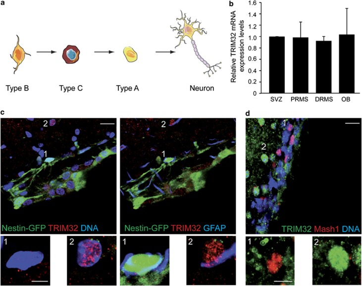
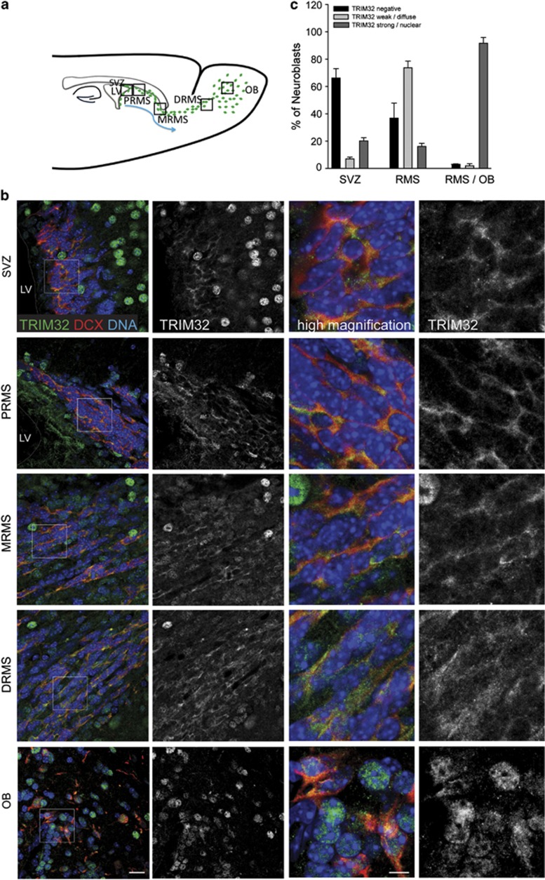
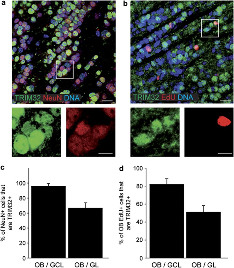
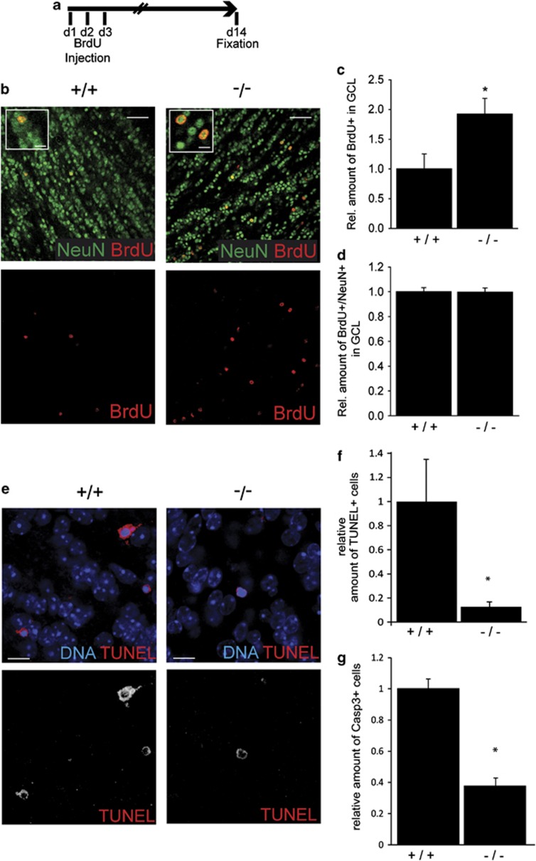
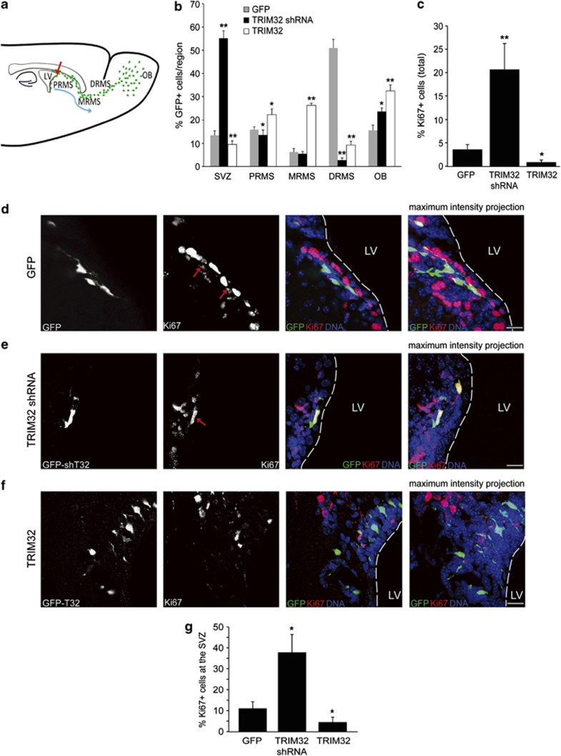
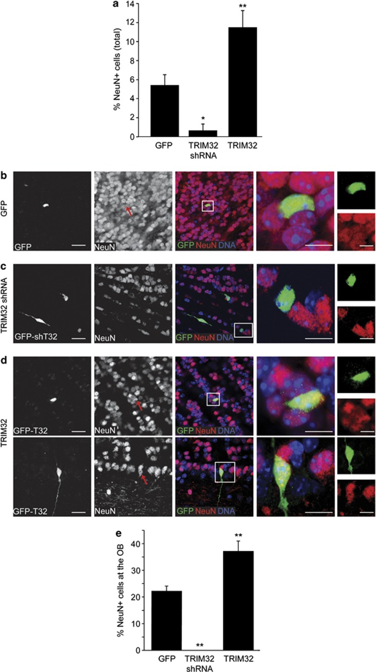
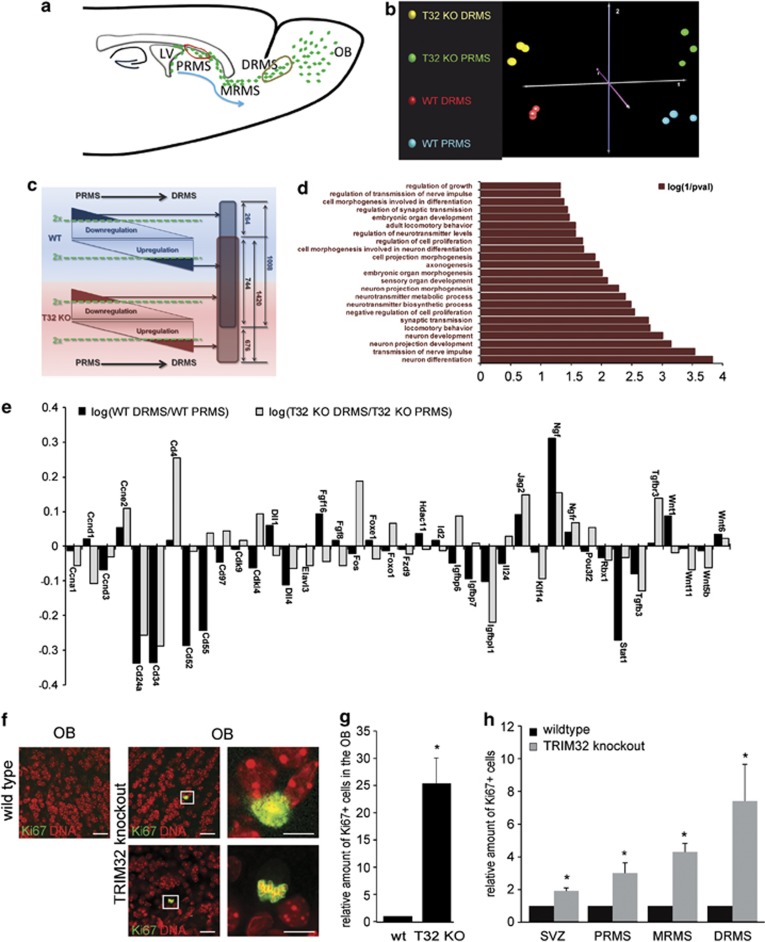
In the adult mammalian brain, neural stem cells in the subventricular zone continuously generate new neurons for the olfactory bulb. Cell fate commitment in these adult neural stem cells is regulated by cell fate-determining proteins. Here, we show that the cell fate-determinant TRIM32 is upregulated during differentiation of adult neural stem cells into olfactory bulb neurons. We further demonstrate that TRIM32 is necessary for the correct induction of neuronal differentiation in these cells. In the absence of TRIM32, neuroblasts differentiate slower and show gene expression profiles that are characteristic of immature cells. Interestingly, TRIM32 deficiency induces more neural progenitor cell proliferation and less cell death. Both effects accumulate in an overproduction of adult-generated olfactory bulb neurons of TRIM32 knockout mice. These results highlight the function of the cell fate-determinant TRIM32 for a balanced activity of the adult neurogenesis process.


Figures

Figure 1
Figure 1
TRIM32 mRNA is expressed throughout the SVZ–OB system, but TRIM32 protein is virtually absent from the majority of type B and C cells. (a) Schematic drawing of the neuronal lineage: type B cells (stem cell astrocytes) in the SVZ give rise to type C cells (transient amplifying cells), which differentiate into type A cells (neuroblasts). Type A cells further differentiate into mature neurons. (b) RT-qPCR measuring the relative TRIM32 mRNA expression levels within the different indicated adult brain regions derived after micro-dissection and Nestin-GFP-based FACS analysis (N=3; mean±S.E.M.; t-test). (c and d) Immunostainings of mouse brain sections labeled with the indicated antibodies. Images were taken in the SVZ. Bars=20 μm, for high magnifications 5 μm (c) and 10 μm (d)
Figure 2
Figure 2
Expression of TRIM32 increases in neuroblasts through the RMS into the OB. (a) Schematic representation of the SVZ–RMS–OB system is shown. (b) Immunostainings of mouse brain sections, taken from regions indicated in panel (a), labeled with the indicated antibodies. Bars=20 μm, high magnification 10 μm. (c) Quantification of the percentage of neuroblasts expressing TRIM32 (mean±S.E.M., t-test, n≥735 cells, N=3 mice). The expression of TRIM32 increases from immature cells in the SVZ to mature neurons in the OB
Figure 3
Figure 3
TRIM32 is strongly expressed in adult-generated neurons of the OB. (a and b) Immunostainings of mouse brain sections, taken in the OB, labeled with the indicated antibodies. Bars=20 μm, for high magnifications 10 μm. (c and d) Diagrams showing the percentage of cells in the GL and GCL of the OB that are double positive for NeuN and TRIM32 (c) and EdU and TRIM32 (d) (mean±S.E.M., n≥3 mice). These results indicate that TRIM32 is expressed in adult-generated neurons of the OB
Figure 4
Figure 4
TRIM32 knockout mice show an increase in the number of newborn neurons and decreased rates of apoptosis. (a) Schematic drawing of the experimental paradigm. For 3 days, mice received a daily injection of BrdU; 14 days later, the brains were fixed and analyzed. (b) Immunostainings of sections from the OB-GCL of wild-type and TRIM32 knockout mice labeled with the indicated antibodies. Bars=50 μm, for high magnifications 15 μm. (cd) Diagrams showing the relative amount of BrdU-positive cells in the OB-GCL (c) and the amount of cells that are double positive for BrdU and NeuN in the OB-GCL (d). All quantifications were conducted in wild-type and knockout mice (mean±S.E.M., N≥4 mice; t-test, * P<0.05). (e) TUNEL stainings of sections from wild-type and TRIM32 knockout mice OB-GCL. Bars=15 μm. (f and g) Diagrams showing the relative amount of TUNEL+ (f) and Casp3+ (g) cells in the OB-GCL. (Median±S.E.M., N≥4 mice, Mann–Whitney U Test *P<0.05). These results show that in the absence of TRIM32 in the adult OB-GCL more cells are generated and that these cells eventually undergo neuronal differentiation
Figure 5
Figure 5
Knockdown of TRIM32 induces proliferation in neural progenitor cells. (a) Schematic representation depicting the migration process of the neural progenitor cells from the SVZ to the OB. Viruses for expression of a scrambled shRNA sequence (control), TRIM32-shRNA or overexpression of TRIM32 were injected in the SVZ. All constructs express GFP under an independent promoter. Eight days after injection, brain sections were analyzed. (b) Quantification of the position of GFP-positive cells in injected brains (mean±S.E.M., N≥700; N>8 mice, Mann–Whitney U test, *P<0.05; **P<0.01). (c) Ki67 was used as a proliferation marker. A quantification of GFP-positive cells that are also positive for Ki67, summarizing all brain regions, is shown (mean±S.E.M., n≥700; N>8 mice, Mann–Whitney U test, *P<0.05; **P<0.01). (df) Immunostainings of mouse brain sections after injection of viruses for GFP, TRIM32-shRNA or TRIM32, taken from the SVZ and labeled with the indicated antibodies. Bars=15 μm. (g) Quantification of GFP-positive cells in the SVZ, which are also positive for Ki67 (mean±S.E.M., n≥500; N≥8 mice, Mann–Whitney U test, *P<0.05; **P<0.01). The results presented here indicate that TRIM32 regulates proliferation of neural progenitor cells
Figure 6
Figure 6
Correct induction of neuronal differentiation depends on the presence of TRIM32. Viruses for expression of a scrambled shRNA sequence (control), TRIM32-shRNA or overexpression of TRIM32 were injected in the SVZ. All constructs express GFP under an independent promoter. Eight days after injection, brain sections were analyzed. (a) NeuN was used as a neuronal differentiation marker. A quantification of GFP-positive cells that are also positive for NeuN, summarizing all brain regions is shown (mean±S.E.M., n≥500; n≥8 mice, Mann–Whitney U test, *P<0.05; **P<0.01). (bd) Immunostainings of mouse brain sections, taken from the OB and labeled with the indicated antibodies. Boxed areas are shown in higher magnification. Red arrows indicate transduced cells that are positive for NeuN. Bars=15 μm for the first three columns, 10 μm for the fourth and fifth columns. (e) Quantification of GFP-positive cells in the OB, which are also positive for NeuN (mean±S.E.M., n≥500; N≥8 mice, Mann–Whitney U test, *P<0.05; **P<0.01). These results indicate that TRIM32 is necessary for the correct induction of neuronal differentiation of neuroblasts
Figure 7
Figure 7
Molecular profiling demonstrates deregulated neuronal differentiation in TRIM32 knockout mice. (a) Schematic representation reflecting the migration and maturation of neural progenitor cells with regions of isolated tissue samples indicated by red (PRMS) and brown (DRMS) circles. (b) CoA performed on the 5000 transcripts with the highest S.D. throughout the series of 12 microarrays analyzed. (c) Schematic illustration of the expression data filtering process. For wild-type (blue) and TRIM32 knockout (red) mice, gene expression levels were compared between the PRMS and the DRMS. Genes with twofold regulation (dark blue or dark red triangles) were isolated for further data processing. In all, 1008 transcripts were at least twofold regulated when comparing PRMS and DRMS in wild-type mice, whereas 1420 transcripts were twofold regulated in TRIM32 knockout animals. From these genes, 744 transcripts were simultaneously designated as regulated in wild-type and TRIM32 knockout mice and thus are varying independently from the genotype. A total of 676 transcripts were exclusively regulated in TRIM32 knockout mice and are therefore specifically associated with loss of TRIM32. (d) GO term analysis of the 676 transcripts exclusively associated with loss of TRIM32. The bar diagram represents the log(1/P-value) (Fisher's exact test). The entire list of GO terms is shown in Supplementary Figure S6. (e) Bar diagram of the expression changes of genes implicated in NSC fate-regulating mechanisms. Black and grey bars represent the log of the ratio calculated from the gene expression intensity within the DRMS and within the PRMS of wild-type and of TRIM32 knockout mice. (f) Immunostainings of brain sections (wild-type and TRIM32 knockout), taken from the OB and labeled with the indicated antibodies. Boxed areas are shown in higher magnification. Bars=25 μm, for high magnifications 10 μm. (g) Bar diagram of the relative amount of Ki67+ cells in the OB of wild-type and TRIM32 knockout mice (mean±S.E.M., N≥4 mice, Mann–Whitney U test, *P<0.05). (h) Bar diagram of the relative amount of Ki67+ cells in the SVZ and RMS of wild-type and TRIM32 knockout mice (mean±S.E.M., N≥4 mice, Mann–Whitney U test, *P<0.05)

Similar articles

Cited by

References

    1. Gotz M, Huttner WB. The cell biology of neurogenesis. Nat Rev Mol Cell Biol. 2005;6:777–788. - PubMed
    1. Gage FH. Mammalian neural stem cells. Science. 2000;287:1433–1438. - PubMed
    1. Zhao C, Deng W, Gage FH. Mechanisms and functional implications of adult neurogenesis. Cell. 2008;132:645–660. - PubMed
    1. Ming GL, Song H. Adult neurogenesis in the mammalian brain: significant answers and significant questions. Neuron. 2011;70:687–702. - PMC - PubMed
    1. Lee CY, Robinson KJ, Doe CQ. Lgl, Pins and aPKC regulate neuroblast self-renewal versus differentiation. Nature. 2006;439:594–598. - PubMed

Publication types

MeSH terms

Substances

LinkOut - more resources