

Spontaneous superior ophthalmic vein thrombosis: a rare entity with potentially devastating consequences
- PMID: 24357838
- PMCID: PMC3965813
- DOI: 10.1038/eye.2013.273
Spontaneous superior ophthalmic vein thrombosis: a rare entity with potentially devastating consequences
Abstract
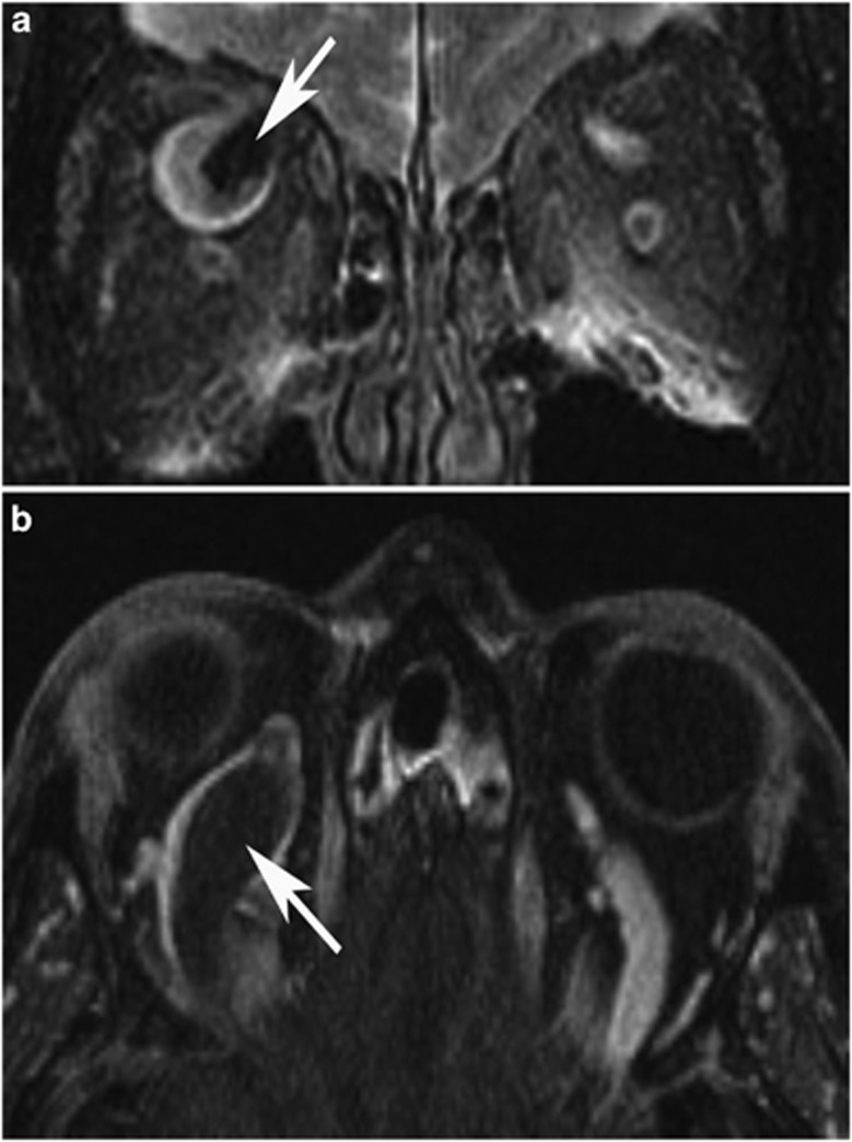
Purpose: Spontaneous superior ophthalmic vein thrombosis (SOVT) is a rare entity. We describe three patients with spontaneous ophthalmic vein thrombosis, each with various risk factors.
Patients and methods: A retrospective review of three patients with a diagnosis of superior ophthalmic vein thrombosis. Clinical characteristics, radiographic features, management techniques and outcomes are described.
Results: All patients presented with unilateral painful proptosis. Two patients had intact light perception, whereas one patient presented with absent light perception. All patients had identifiable risk factors for thrombosis, which included sickle cell trait, hereditary hemorrhagic telangectasia and colon cancer with recurrent deep vein thrombosis. Anticoagulation was initiated in two patients. Resolution of proptosis was seen in all patients, with no recovery of vision in one patient.
Conclusions: Risk factors for spontaneous superior ophthalmic vein thrombosis are multifactorial. MRI and MRV confirm the diagnosis of SOVT. Despite urgent intervention devastating visual loss may occur.
Figures


References
-
- Stiebel-Kalish H, Setton A, Nimii Y, Kalish Y, Hartman J, Huna Bar-On R, et al. Cavernous sinus dural arteriovenous malformations: patterns of venous drainage are related to clinical signs and symptoms. Ophthalmology. 2002;109:1685–1691. - PubMed
-
- Schmitt NJ, Beatty RL, Kennerdell JS. Superior ophthalmic vein thrombosis in a patient with dacryocystitis-induced orbital cellulitis. Ophthal Plast Reconstr Surg. 2005;21:387–389. - PubMed
-
- Shinder R, Oellers P, Esmaeli B, Schiffman JS. Superior ophthalmic vein thrombosis in a patient with chronic myeloid leukemia receiving antifibrinolytic and thrombopoietin receptor agonist therapy. J Ocul Pharmacol Ther. 2010;26:293–296. - PubMed
-
- Shovlin CL, Guttmacher AE, Buscarini E, Faughnan ME, Hyland RH, Westermann CJ, et al. Diagnostic criteria for hereditary hemorrhagic telangiectasia (Rendu-Osler-Weber syndrome) Am J Med Genet. 2000;91:66–67. - PubMed
-
- Bagot CN, Arya R. Virchow and his triad: a question of attribution. Br J Haematol. 2008;143:180–190. - PubMed
Publication types
MeSH terms
Substances
LinkOut - more resources
Full Text Sources
Other Literature Sources
Medical
