

Imaging Early Demineralization on Tooth Occlusal Surfaces with a High Definition InGaAs Camera
- PMID: 24357911
- PMCID: PMC3865214
- DOI: 10.1117/12.2011015
Imaging Early Demineralization on Tooth Occlusal Surfaces with a High Definition InGaAs Camera
Abstract
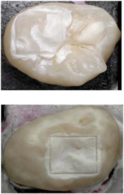
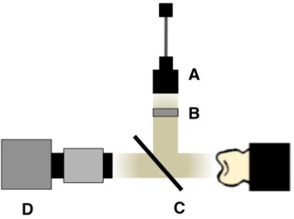
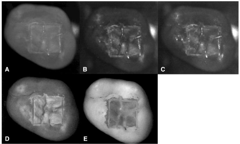
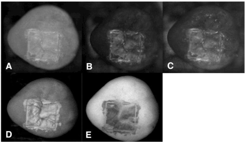
In vivo and in vitro studies have shown that high contrast images of tooth demineralization can be acquired in the near-IR due to the high transparency of dental enamel. The purpose of this study is to compare the lesion contrast in reflectance at near-IR wavelengths coincident with high water absorption with those in the visible, the near-IR at 1300-nm and with fluorescence measurements for early lesions in occlusal surfaces. Twenty-four human molars were used in this in vitro study. Teeth were painted with an acid-resistant varnish, leaving a 4×4 mm window in the occlusal surface of each tooth exposed for demineralization. Artificial lesions were produced in the exposed windows after 1 & 2-day exposure to a demineralizing solution at pH 4.5. Lesions were imaged using NIR reflectance at 3 wavelengths, 1310, 1460 and 1600-nm using a high definition InGaAs camera. Visible light reflectance, and fluorescence with 405-nm excitation and detection at wavelengths greater than 500-nm were also used to acquire images for comparison. Crossed polarizers were used for reflectance measurements to reduce interference from specular reflectance. The contrast of both the 24 hr and 48 hr lesions were significantly higher (P<0.05) for NIR reflectance imaging at 1460-nm and 1600-nm than it was for NIR reflectance imaging at 1300-nm, visible reflectance imaging, and fluorescence. The results of this study suggest that NIR reflectance measurements at longer near-IR wavelengths coincident with higher water absorption are better suited for imaging early caries lesions.
Keywords: Near-IR imaging; artificial lesions; demineralization; dental caries; enamel; polarization.
Figures
Similar articles
-
High contrast reflectance imaging of simulated lesions on tooth occlusal surfaces at near-IR wavelengths.Lasers Surg Med. 2013 Oct;45(8):533-41. doi: 10.1002/lsm.22159. Epub 2013 Jul 16. Lasers Surg Med. 2013. PMID: 23857066 Free PMC article.
-
Multispectral near-IR reflectance imaging of simulated early occlusal lesions: variation of lesion contrast with lesion depth and severity.Lasers Surg Med. 2014 Mar;46(3):203-15. doi: 10.1002/lsm.22216. Epub 2013 Dec 27. Lasers Surg Med. 2014. PMID: 24375543 Free PMC article.
-
High contrast near-infrared polarized reflectance images of demineralization on tooth buccal and occlusal surfaces at lambda = 1310-nm.Lasers Surg Med. 2009 Mar;41(3):208-13. doi: 10.1002/lsm.20746. Lasers Surg Med. 2009. PMID: 19291753 Free PMC article.
-
Multispectral near-IR reflectance and transillumination imaging of teeth.Biomed Opt Express. 2011 Oct 1;2(10):2804-14. doi: 10.1364/BOE.2.002804. Epub 2011 Sep 15. Biomed Opt Express. 2011. PMID: 22025986 Free PMC article.
-
Near-IR and CP-OCT imaging of suspected occlusal caries lesions.Lasers Surg Med. 2017 Mar;49(3):215-224. doi: 10.1002/lsm.22641. Epub 2017 Mar 24. Lasers Surg Med. 2017. PMID: 28339115 Free PMC article.
Cited by
-
Comparative Diagnostic Accuracy of VistaCam IX Proxi and Bitewing Radiography for Detection of Interproximal Caries.J Dent (Shiraz). 2023 Dec 1;24(4):395-403. doi: 10.30476/dentjods.2022.95326.1860. eCollection 2023 Dec. J Dent (Shiraz). 2023. PMID: 38149228 Free PMC article.
-
Near-infrared fluorescence imaging in the largely unexplored window of 900-1,000 nm.Theranostics. 2018 Jul 16;8(15):4116-4128. doi: 10.7150/thno.26539. eCollection 2018. Theranostics. 2018. PMID: 30128040 Free PMC article.
-
Near-Infrared Imaging for Detecting Caries and Structural Deformities in Teeth.IEEE J Transl Eng Health Med. 2017 Apr 19;5:2300107. doi: 10.1109/JTEHM.2017.2695194. eCollection 2017. IEEE J Transl Eng Health Med. 2017. PMID: 28507826 Free PMC article.
References
-
- NIH Diagnosis and Management of Dental Caries throughout Life. NIH Consensus Statement. 2001:1–24. - PubMed
-
- Featherstone JDB. Prevention and reversal of dental caries:role of low level fluoride. Community Dent Oral Epidemiol. 1999;27:31–40. - PubMed
-
- Angmar-Mansson B, ten Bosch JJ. Optical methods for the detection and quantification of caries. Adv. Dent. Res. 1987;1(1):14–20. - PubMed
-
- ten Bosch JJ, van der Mei HC, Borsboom PCF. Optical monitor of in vitro caries. Caries Res. 1984;18:540–547. - PubMed
-
- Benson PE, Ali Shah A, Robert Willmot D. Polarized versus nonpolarized digital images for the measurement of demineralization surrounding orthodontic brackets. Angle Orthod. 2008;78(2):288–293. - PubMed
Grants and funding
LinkOut - more resources
Full Text Sources
Other Literature Sources
Miscellaneous