

Regeneration of the corneal epithelium with conjunctival epithelial equivalents generated in serum- and feeder-cell-free media
- PMID: 24357922
- PMCID: PMC3867160
Regeneration of the corneal epithelium with conjunctival epithelial equivalents generated in serum- and feeder-cell-free media
Abstract
Purpose: An alternative autologous tissue for ocular surface reconstruction is a potential treatment for the patients with bilateral limbal stem cell deficiency. For the purpose of regenerative procedures in patients, it is desirable to eliminate the involvement of xenogeneic components, such as nonhuman sera and feeder cells. In the present study, we examined the behavior and phenotypic features of cultured conjunctival epithelial sheets generated in serum- and 3T3-free culture conditions when transplanted into the de-epithelialized limbal corneal surface.
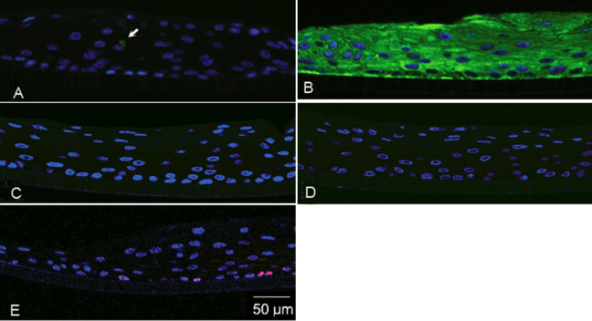
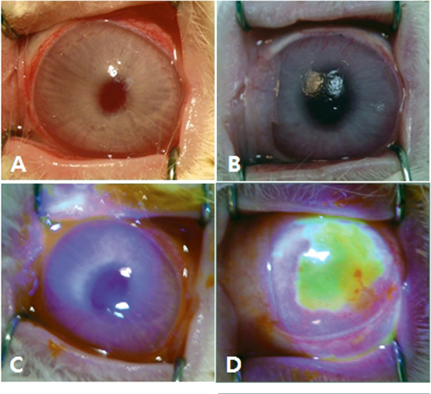
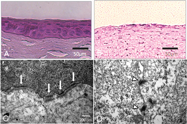
Methods: Epithelial cells from normal conjunctiva obtained by neutral protease digestion were expanded by culture in a serum-free low-calcium medium and set in an air-liquid interface culture for 14 days. The resulting multilayered epithelial sheets were grafted onto rabbit ocular surfaces made epithelial-free by alkali treatment. Pre-grafted and post-grafted epithelia were analyzed by electron microscopy and immunohistochemistry.
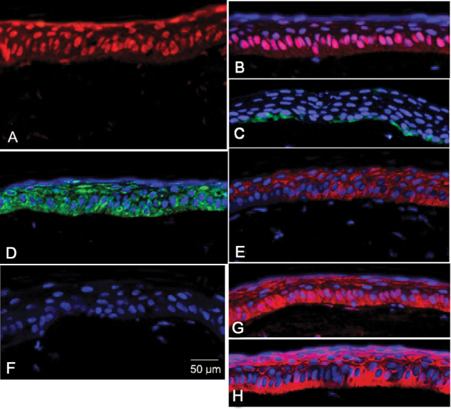
Results: At graft time the cultured epithelial sheet consisted of 6-8 layers of properly stratified epithelium that displayed a CK19(+)/MUC5AC(+)/ CK3 (-)/CK12(-) phenotype, consistent with the conjunctival epithelial lineage. Two weeks after xeno-grafting the in vivo epithelium consisted of 5-6 well compacted layers expressing the precursor cell-related protein p63, the proliferation marker Ki67, desmosomes, hemidesmosomes and its integrin (β4), and the corneal specific cytokeratins CK3, and CK12. Conjunctival goblet cell mucin (MUC5AC) was not visible. The engrafted epithelium stained positively for the anti-human nuclei antibody, confirming that the epithelial cells on the rabbit corneas were of human origin.
Conclusions: Our results suggest that conjunctival epithelial sheets generated in serum- and 3T3-free culture conditions can acquire the corneal epithelial phenotype when transferred to the in vivo corneal stromal environment.
Figures
References
-
- Tsai RJ, Sun TT, Tseng SC. Comparison of limbal and conjunctival autograft transplantation in corneal surface reconstruction in rabbits. Ophthalmology. 1990;97:446–55. - PubMed
-
- Pellegrini G, Traverso CE, Franzi AT, Zingirian M, Cancedda R, De Luca M. Long-term restoration of damaged corneal surfaces with autologous cultivated corneal epithelium. Lancet. 1997;349:990–3. - PubMed
-
- Tsubota K, Satake Y, Kaido M, Shinozaki N, Shimmura S, Bissen-Miyajima H, Shimazaki J. Treatment of severe ocular-surface disorders with corneal epithelial stem-cell transplantation. N Engl J Med. 1999;340:1697–703. - PubMed
-
- Koizumi N, Inatomi T, Suzuki T, Sotozono C, Kinoshita S. Cultivated corneal epithelial transplantation for ocular surface reconstruction in acute phase of Stevens-Johnson syndrome. Arch Ophthalmol. 2001;119:298–300. - PubMed
-
- Sangwan VS, Basu S, Vemuganti GK, Sejpal K, Subramaniam SV, Bandyopadhyay S, Krishnaiah S, Gaddipati S, Tiwari S, Balasubramanian D. Clinical outcomes of xeno-free autologous cultivated limbal epithelial transplantation: a 10-year study. Br J Ophthalmol. 2011;95:1525–9. - PubMed
Publication types
MeSH terms
Substances
Grants and funding
LinkOut - more resources
Full Text Sources
Other Literature Sources
Miscellaneous