

Interaction network of proteins associated with human cytomegalovirus IE2-p86 protein during infection: a proteomic analysis
- PMID: 24358118
- PMCID: PMC3864812
- DOI: 10.1371/journal.pone.0081583
Interaction network of proteins associated with human cytomegalovirus IE2-p86 protein during infection: a proteomic analysis
Abstract
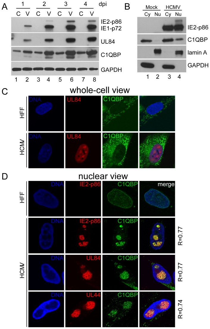
Human cytomegalovirus protein IE2-p86 exerts its functions through interaction with other viral and cellular proteins. To further delineate its protein interaction network, we generated a recombinant virus expressing SG-tagged IE2-p86 and used tandem affinity purification coupled with mass spectrometry. A total of 9 viral proteins and 75 cellular proteins were found to associate with IE2-p86 protein during the first 48 hours of infection. The protein profile at 8, 24, and 48 h post infection revealed that UL84 tightly associated with IE2-p86, and more viral and cellular proteins came into association with IE2-p86 with the progression of virus infection. A computational analysis of the protein-protein interaction network indicated that all of the 9 viral proteins and most of the cellular proteins identified in the study are interconnected to varying degrees. Of the cellular proteins that were confirmed to associate with IE2-p86 by immunoprecipitation, C1QBP was further shown to be upregulated by HCMV infection and colocalized with IE2-p86, UL84 and UL44 in the virus replication compartment of the nucleus. The IE2-p86 interactome network demonstrated the temporal development of stable and abundant protein complexes that associate with IE2-p86 and provided a framework to benefit future studies of various protein complexes during HCMV infection.
Conflict of interest statement
Figures


Similar articles
-
Characteristics of Immediate-Early 2 (IE2) and UL84 Proteins in UL84-Independent Strains of Human Cytomegalovirus (HCMV).Microbiol Spectr. 2021 Oct 31;9(2):e0053921. doi: 10.1128/Spectrum.00539-21. Epub 2021 Sep 22. Microbiol Spectr. 2021. PMID: 34550009 Free PMC article.
-
Covalent modification of the transactivator protein IE2-p86 of human cytomegalovirus by conjugation to the ubiquitin-homologous proteins SUMO-1 and hSMT3b.J Virol. 2000 Mar;74(6):2510-24. doi: 10.1128/jvi.74.6.2510-2524.2000. J Virol. 2000. PMID: 10684265 Free PMC article.
-
The UL84 protein of human cytomegalovirus acts as a transdominant inhibitor of immediate-early-mediated transactivation that is able to prevent viral replication.J Virol. 1997 Sep;71(9):7048-60. doi: 10.1128/JVI.71.9.7048-7060.1997. J Virol. 1997. PMID: 9261435 Free PMC article.
-
Identification of additional IE2-p86-responsive cis-repressive sequences within the human cytomegalovirus major immediate early gene promoter.J Biomed Sci. 2002 Sep-Oct;9(5):460-70. doi: 10.1007/BF02256541. J Biomed Sci. 2002. PMID: 12218362
-
Human cytomegalovirus UL84 oligomerization and heterodimerization domains act as transdominant inhibitors of oriLyt-dependent DNA replication: evidence that IE2-UL84 and UL84-UL84 interactions are required for lytic DNA replication.J Virol. 2004 Sep;78(17):9203-14. doi: 10.1128/JVI.78.17.9203-9214.2004. J Virol. 2004. PMID: 15308715 Free PMC article.
Cited by
-
The Interactions of the Complement System with Human Cytomegalovirus.Viruses. 2024 Jul 20;16(7):1171. doi: 10.3390/v16071171. Viruses. 2024. PMID: 39066333 Free PMC article. Review.
-
Characteristics of Immediate-Early 2 (IE2) and UL84 Proteins in UL84-Independent Strains of Human Cytomegalovirus (HCMV).Microbiol Spectr. 2021 Oct 31;9(2):e0053921. doi: 10.1128/Spectrum.00539-21. Epub 2021 Sep 22. Microbiol Spectr. 2021. PMID: 34550009 Free PMC article.
-
The life cycle and pathogenesis of human cytomegalovirus infection: lessons from proteomics.Expert Rev Proteomics. 2014 Dec;11(6):697-711. doi: 10.1586/14789450.2014.971116. Epub 2014 Oct 18. Expert Rev Proteomics. 2014. PMID: 25327590 Free PMC article.
-
Interaction of human cytomegalovirus pUL52 with major components of the viral DNA encapsidation network underlines its essential role in genome cleavage-packaging.J Virol. 2025 Apr 15;99(4):e0220124. doi: 10.1128/jvi.02201-24. Epub 2025 Mar 10. J Virol. 2025. PMID: 40062846 Free PMC article.
-
Hsp70 Isoforms Are Essential for the Formation of Kaposi's Sarcoma-Associated Herpesvirus Replication and Transcription Compartments.PLoS Pathog. 2015 Nov 20;11(11):e1005274. doi: 10.1371/journal.ppat.1005274. eCollection 2015 Nov. PLoS Pathog. 2015. PMID: 26587836 Free PMC article.
References
-
- Mocarski ES, Shenk T, Pass RF (2007) cytomegalovirus. In D MKnipe and P M Howley (ed), Fields Virology Lippincott Williams & Wilkins, Philadelphia, PA.
Publication types
MeSH terms
Substances
Grants and funding
LinkOut - more resources
Full Text Sources
Other Literature Sources
Medical
Miscellaneous
