. 2013 Dec 16;8(12):e81774.
doi: 10.1371/journal.pone.0081774. eCollection 2013.

IL-4/IL-13-dependent and independent expression of miR-124 and its contribution to M2 phenotype of monocytic cells in normal conditions and during allergic inflammation

Affiliations

IL-4/IL-13-dependent and independent expression of miR-124 and its contribution to M2 phenotype of monocytic cells in normal conditions and during allergic inflammation

Tatyana Veremeyko et al. PLoS One. .

Abstract

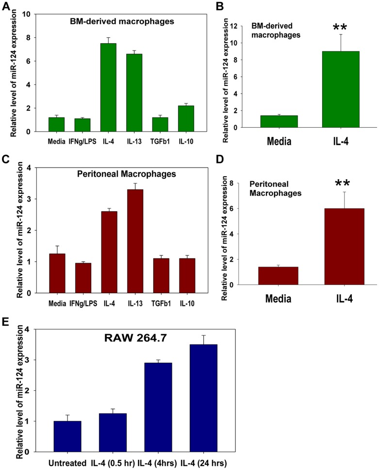
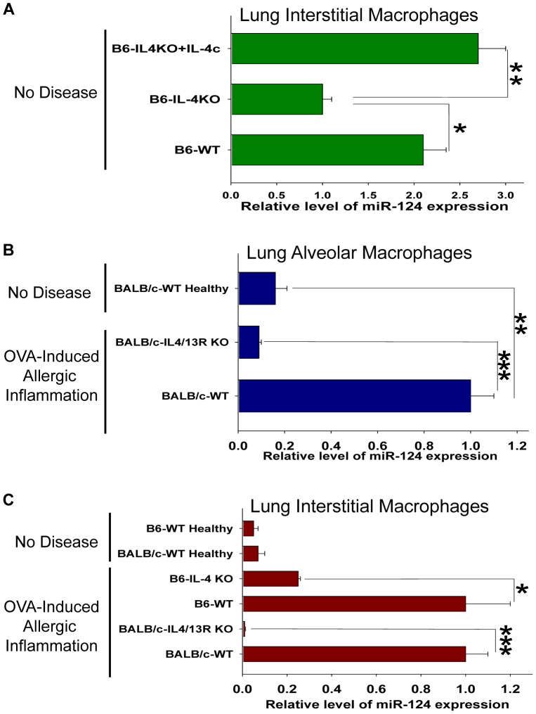
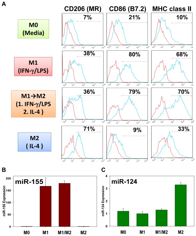
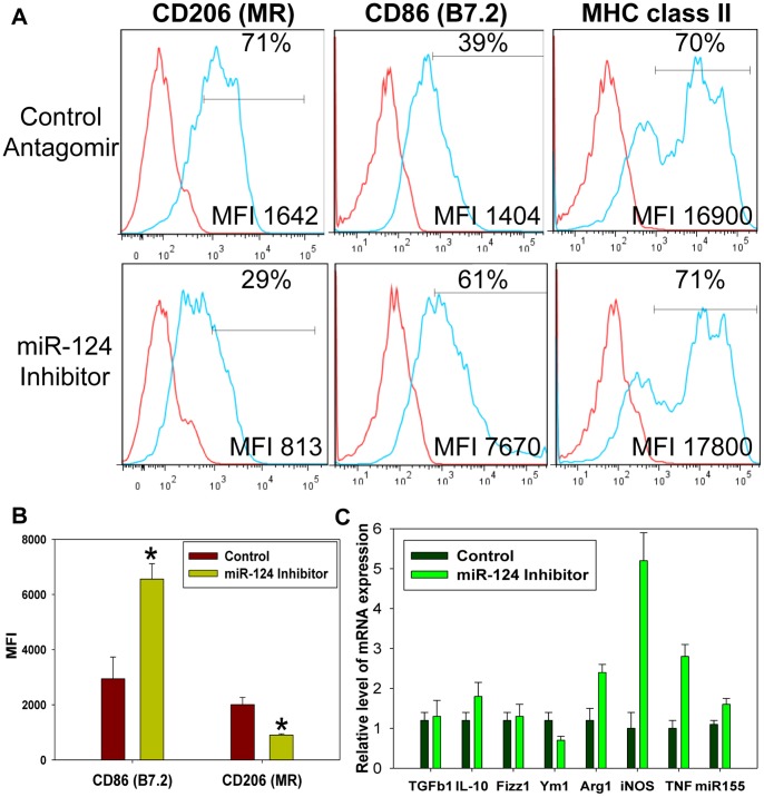
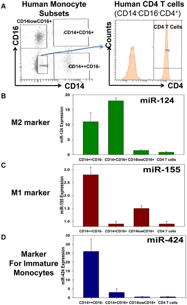
Monocytic cells exhibit a high level of heterogeneity and have two distinct modes of their activation: 1) classical M1 path associated with inflammation and tissue damage, and 2) alternative M2 path. Although it has been demonstrated that M2 macrophages play an important role in the regulation of the allergic immune responses, tissue maintenance and repair, little is known about the mechanisms that determine the M2 phenotype. We have previously shown that miR-124 is expressed in microglia that exhibit the M2 phenotype and overexpression of miR-124 in macrophages resulted in downregulation of a number of M1 markers (MHC class II, CD86) and up-regulation of several M2 markers (Fizz1, Arg1). We further investigated whether the polarization of macrophages towards the M2 phenotype induced miR-124 expression. We found that exposure of cells to IL-4 and IL-13 resulted in the upregulation of miR-124 in macrophages. We also demonstrated that IL-4 induced expression of three miR-124 precursor transcripts with predominant expression of pri-miR-124.3, suggesting regulation of miR-124 expression by IL-4 on a transcriptional level. Expression of miR-124 in microglia did not depend on IL-4 and/or IL-13, whereas expression of miR-124 in lung resident macrophages was IL-4 and IL-13-dependent and was upregulated by systemic administration of IL-4 or during allergic inflammation. Upregulation of several M2 markers (CD206, Ym1) and downregulation of the M1 markers (CD86, iNOS, TNF) in M2-polarized macrophages was abrogated by a miR-124 inhibitor, suggesting that this microRNA contributed to the M2 phenotype development and maintenance. Finally we showed that human CD14(+)CD16(+) intermediate monocytes, which are found in increased numbers in patients with allergies and bronchial asthma, expressed high levels of miR-124 and exhibited other properties of M2-like cells. Thus, our study suggests that miR-124 serves as a regulator of the M2 polarization in various subsets of monocytic cells both in vitro and in vivo.


Conflict of interest statement

Competing Interests: The authors have declared that no competing interests exist.

Figures

Figure 1
Figure 1. Analysis of miR-124 expression in macrophages polarized in vitro towards M1 (IFN-γ/LPS), M2a (IL-4, or IL-13), or M2b (TGF-β1, or IL-10).
Bone marrow (BM) derived cultured macrophages (A, B), ex-vivo isolated CD11b+F4/80+ peritoneal macrophages (C, D), or macrophage cell line RAW 264.7 (E) were incubated with media alone (Media), or IFN-γ and LPS (IFN-γ/LPS), or IL-4, or IL-13, or TGF-β1, or IL-10 for 24 hours, after which the expression of miR-124 was measured by the real-time RT-PCR as described in Materials and Methods. The data is representative of eight (A, B), six (C, D) or five (E) separate experiments; mean ± S.E. of triplicate is shown in (A, C, E). Mean ± S.E. of eight separate experiments is shown in (B) and mean ± S.E. of six separate experiments is shown in (D) (**p<0.01).
Figure 2
Figure 2. Analysis of the miR-124 expression in lung macrophages in the baseline conditions or during the OVA-induced allergic lung inflammation.
(A) Lung interstitial macrophages were isolated from the unmanipulated B6-WT mice, B6-IL-4 deficient mice, or B6-IL-4 deficient mice in which IL-4c was systemically administered as described in Materials and Methods and the expression of miR-124 was measured as in Fig. 1. (B) Lung alveolar macrophages were isolated from the unmanipulated BALB/c-WT healthy mice, or BALB/c-WT and BALB/c-IL-4/13R deficient mice with OVA-induced allergic lung inflammation and the expression of miR-124 was analyzed. (C) Lung interstitial macrophages were isolated from healthy B6-WT and healthy BALB/c-WT vs. B6-WT and B6-IL-4 deficient mice, or BALB/c-WT and BALB/c-IL-4/13R deficient mice with an OVA-induced allergic lung inflammation and expression of miR-124 was analyzed. In (A–C), Mean ± S.E. of 4–6 individual animals is shown (*, p<0.05; **, p<0.01; ***, p<0.005).
Figure 3
Figure 3. Treatment of M1 macrophages with IL-4 did not result in upregulation of miR-124 and conversion of M1 cells into M2.
Peritoneal CD11b+F4/80+ macrophages were isolated and left unpolarized (M0) or polarized towards M1, or M2, or first polarized towards M1, and after 24 hours, washed and polarized towards M2 (M1→M2 or M/M2) as described in Materials and Methods. After which, the cells were stained for CD206, CD86 and MHC class II and analyzed by FACS (A), or used for RNA isolation and analysis of expression of miR-155 (B) or miR-124 (C) by real-time RT-PCR. One representative experiment of three is shown in (A–C); mean ± S.E. of triplicate is shown in (B, C).
Figure 4
Figure 4. miR-124 inhibitor decreased expression of M2 markers CD206 and Ym-1 and increased expression of M1 markers CD86, iNOS, TNF, and miR-155 in the in-vitro polarized M2 macrophages.
Peritoneal CD11b+F4/80+ macrophages were isolated and treated with IL-4 in the presence of liposomes with miR-124 inhibitor or control antagomir for 24 hours as described in Materials and Methods. After which, viable cells were separated using Ficoll gradient and either stained for CD206, CD86 and MHC class II and analyzed by FACS (A, B), or used for mRNA isolation and analysis of expression of mRNA transcripts for TGF-β1, IL-10, Fizz1, Ym1, Arg1, iNOS, TNF, and miR-155 (C). In (A), the red line represents staining for isotype-matched control and the blue line - staining for surface markers. Each histogram shows overlay images. The percentage of positive cells is shown on each histogram in the upper right corner and the mean fluorescence intensity (MFI) - in the bottom right corner. The mean MFI ± S.E. of three separate experiments is shown in (B) (*, p<0.05) and mean ± S.E. of a triplicate is shown in (C).
Figure 5
Figure 5. Analysis of expression of miR-124 (the M2 marker), miR-155 (the M1 marker) and miR-424 (the marker for immature monocytes) in the populations of classical (CD14++CD16
), intermediate (CD14+CD16+) and non-classical (CD14lowCD16+) human monocytes. (A) Mononuclear cells were isolated from the peripheral blood of healthy individuals and stained for CD14, CD16 and CD4. The populations of CD14++CD16, CD14+CD16+, and CD14lowCD16+ monocytes and CD14-CD16-CD4+ CD4 subsets were sorted by FACS and the expression of miR-124, miR-155, and miR-424 was analyzed as described in Materials and Methods. The mean MFI ± S.E. of three separate experiments with three healthy individuals is shown. Expression of miR-124 (B), miR-155 (C) and miR-424 (D) was analyzed in three sorted monocyte populations and compared to CD4 T cells. The level of expression of indicated miRNAs in CD4 T cells was used as a reference value.

References

    1. Wynn TA, Chawla A, Pollard JW (2013) Macrophage biology in development, homeostasis and disease. Nature 496: 445–455. - PMC - PubMed
    1. Geissmann F, Manz MG, Jung S, Sieweke MH, Merad M, et al. (2010) Development of monocytes, macrophages, and dendritic cells. Science 327: 656–661. - PMC - PubMed
    1. Jenkins SJ, Ruckerl D, Cook PC, Jones LH, Finkelman FD, et al. (2011) Local macrophage proliferation, rather than recruitment from the blood, is a signature of TH2 inflammation. Science 332: 1284–1288. - PMC - PubMed
    1. Gordon S, Taylor PR (2005) Monocyte and macrophage heterogeneity. Nat Rev Immunol 5: 953–964. - PubMed
    1. Zhao C, Zhang H, Wong WC, Sem X, Han H, et al. (2009) Identification of novel functional differences in monocyte subsets using proteomic and transcriptomic methods. J Proteome Res 8: 4028–4038. - PubMed

Publication types

MeSH terms