. 2013 Dec 16;8(12):e82285.
doi: 10.1371/journal.pone.0082285. eCollection 2013.

Identification of crucial amino acids in mouse aldehyde oxidase 3 that determine substrate specificity

Affiliations

Identification of crucial amino acids in mouse aldehyde oxidase 3 that determine substrate specificity

Martin Mahro et al. PLoS One. .

Abstract

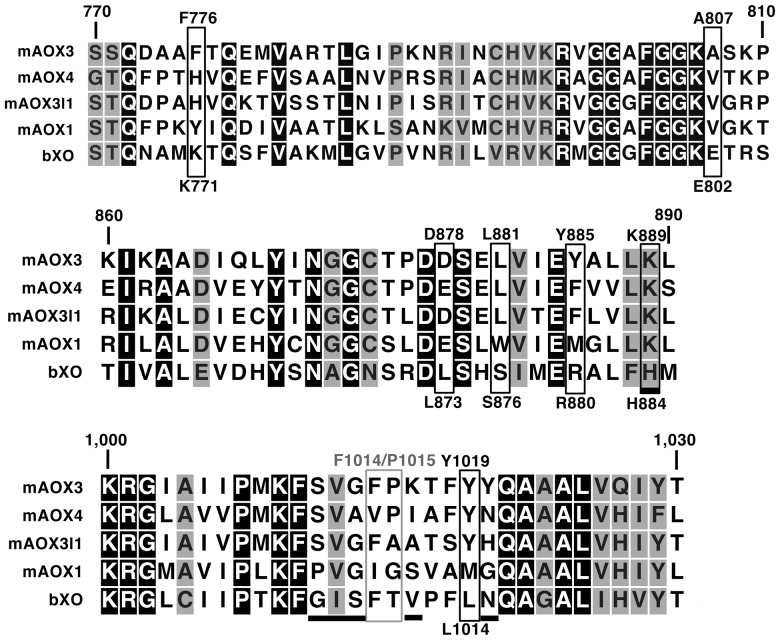
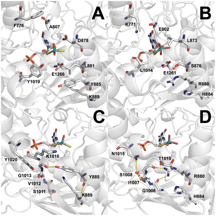
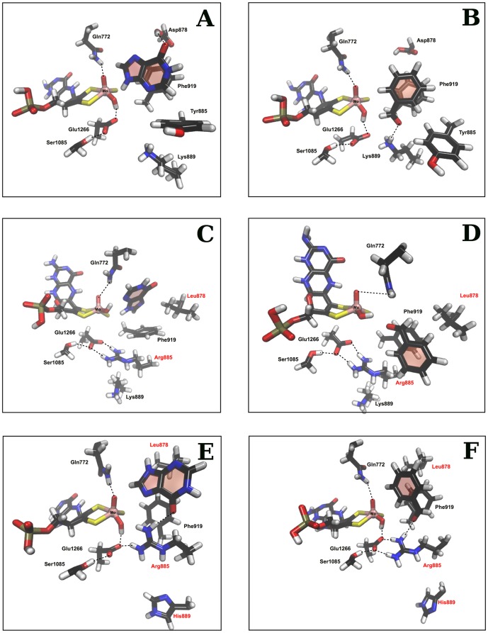
In order to elucidate factors that determine substrate specificity and activity of mammalian molybdo-flavoproteins we performed site directed mutagenesis of mouse aldehyde oxidase 3 (mAOX3). The sequence alignment of different aldehyde oxidase (AOX) isoforms identified variations in the active site of mAOX3 in comparison to other AOX proteins and xanthine oxidoreductases (XOR). Based on the structural alignment of mAOX3 and bovine XOR, differences in amino acid residues involved in substrate binding in XORs in comparison to AOXs were identified. We exchanged several residues in the active site to the ones found in other AOX homologues in mouse or to residues present in bovine XOR in order to examine their influence on substrate selectivity and catalytic activity. Additionally we analyzed the influence of the [2Fe-2S] domains of mAOX3 on its kinetic properties and cofactor saturation. We applied UV-VIS and EPR monitored redox-titrations to determine the redox potentials of wild type mAOX3 and mAOX3 variants containing the iron-sulfur centers of mAOX1. In addition, a combination of molecular docking and molecular dynamic simulations (MD) was used to investigate factors that modulate the substrate specificity and activity of wild type and AOX variants. The successful conversion of an AOX enzyme to an XOR enzyme was achieved exchanging eight residues in the active site of mAOX3. It was observed that the absence of the K889H exchange substantially decreased the activity of the enzyme towards all substrates analyzed, revealing that this residue has an important role in catalysis.


Conflict of interest statement

Competing Interests: The authors have declared that no competing interests exist.

Figures

Figure 1
Figure 1. Amino acid sequence alignment of selected parts of mAOX homologues and bovine XOR.
Shown is an amino acid sequence alignment of the region of amino acids 770-1030 of mAOX3 with the other AOX-isoform from mouse and bovine XOR (bXOR). Identical amino acid residues are shaded in black, homologous amino acid residues are shaded in grey. For clarity, residues exchanged in mAOX3-“active site1”-K889H variant are boxed in black. A grey box marks the position of mAOX3-F1014 and mAOX3-P1015 variants. The six residues exchanged in the mAOX3-“gating loop” variants are underlined. The alignment was created with CLC Sequence viewer Ver. 6.8.2.
Figure 2
Figure 2. 8% native-PAGE of mAOX3 wild type and variants.
Lanes 1–12 contain 3–5 µg of purified protein, stained with Coomassie-brilliant blue. 1, mAOX3 WT; 2, mAOX3-“active site1”; 3, mAOX3-“active site1”-K889H; 4, mAOX3-“active site1 + gating loop”; 5, mAOX3-“gating loop”; 6, mAOX3-FeSI-mAOX1; 7, mAOX3-FeSII-mAOX1; 8, mAOX3-FeSII-FeSI-mAOX1; 9, mAOX3-P1015A; 10, mAOX3-P1015G; 11, mAOX3-F1013I; 12, mAOX3-F1014V.
Figure 3
Figure 3. Active site structure of mAOX3.
A, Stick representation of E1266 and residues exchanged in mAOX3-“active site1”-K889H in the crystal structure of mAOX3 WT (pdb:3ZYV). B, Stick representation of residues of desulfurated bXO (pdb:3EUB) corresponding to the amino acids in Panel A. C, Stick representation of residues in mAOX3 corresponding to the residues shown in Panel D. Y885 builds hydrogen-bonds to the backbone of G1013 and K889, indicated by yellow dotted lines D, Stick representation of residues involved in a hydrogen network at the entrance to the active site of bXOR. The hydrogen-bonding network is established by R880, G1006, I1007, S1008 and N1015 in bXOR represented by yellow dotted lines. Through the interactions, the position and orientation of T1010 is altered in comparison to K1016 of mAOX3 in panel C. Figures were created using MacPymol Ver. 0.99rc6.
Figure 4
Figure 4. Molecular docking and molecular dynamic studies.
A, Wild type enzyme with hypoxanthine. B, Wild type enzyme with benzaldehyde bound in the active site. C, mAOX3-“active site1” variant with hypoxanthine. D, mAOX3-“active site1” variant with benzaldehyde bound in the active site. E, mAOX3-“active site1”-K889H with hypoxanthine. F, mAOX3-“active site1”-K889H with benzaldehyde bound in the active site. Hydrogen bonds are marked with a dashed line and the altered residues are highlighted in red.

References

    1. Krenitsky TA, Neil SM, Elion GB, Hitchings GH (1972) A comparison of the specificities of xanthine oxidase and aldehyde oxidase. Arch Biochem Biophys 150: 585–599. - PubMed
    1. Kitamura S, Sugihara K, Ohta S (2006) Drug-metabolizing ability of molybdenum hydroxylases. Drug metabolism and pharmacokinetics 21: 83–98. - PubMed
    1. Garattini E, Mendel R, Romao MJ, Wright R, Terao M (2003) Mammalian molybdo-flavoenzymes, an expanding family of proteins: structure, genetics, regulation, function and pathophysiology. The Biochemical journal 372: 15–32. - PMC - PubMed
    1. Johnson JL, Chaudhury M, Rajagopalan KV (1991) Identification of a molybdopterin-containing molybdenum cofactor in xanthine dehydrogenase from Pseudomonas aeruginosa. Biofactors 3: 103–107. - PubMed
    1. Wahl RC, Rajagopalan KV (1982) Evidence for the inorganic nature of the cyanolyzable sulfur of molybdenum hydroxylases. The Journal of biological chemistry 257: 1354–1359. - PubMed

Publication types

Substances

LinkOut - more resources