

Role of endothelial progenitor cells and inflammatory cytokines in healing of diabetic foot ulcers
- PMID: 24358275
- PMCID: PMC3865213
- DOI: 10.1371/journal.pone.0083314
Role of endothelial progenitor cells and inflammatory cytokines in healing of diabetic foot ulcers
Abstract
Background: To evaluate changes in endothelial progenitor cells (EPCs) and cytokines in patients with diabetic foot ulceration (DFU) in association with wound healing.
Methods: We studied healthy subjects, diabetic patients not at risk of DFU, at risk of DFU and with active DFU. We prospectively followed the DFU patients over a 12-week period. We also investigated similar changes in diabetic rabbit and mouse models of wound healing.
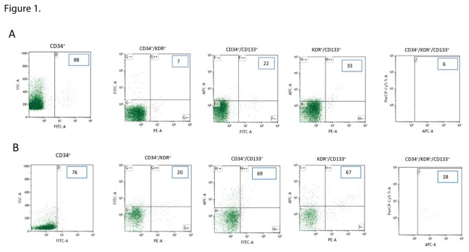
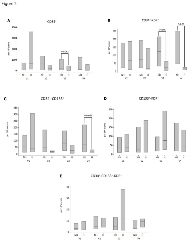
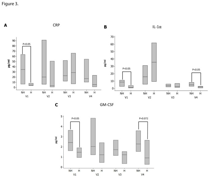
Results: All EPC phenotypes except the kinase insert domain receptor (KDR)(+)CD133(+) were reduced in the at risk and the DFU groups compared to the controls. There were no major EPC differences between the control and not at risk group, and between the at risk and DFU groups. Serum stromal-cell derived factor-1 (SDF-1) and stem cell factor (SCF) were increased in DFU patients. DFU patients who healed their ulcers had lower CD34(+)KDR(+) count at visits 3 and 4, serum c-reactive protein (CRP) and granulocyte-macrophage colony-stimulating factor (GM-CSF) at visit 1, interleukin-1 (IL-1) at visits 1 and 4. EPCs tended to be higher in both diabetic animal models when compared to their non-diabetic counterparts both before and ten days after wounding.
Conclusions: Uncomplicated diabetes does not affect EPCs. EPCs are reduced in patients at risk or with DFU while complete wound healing is associated with CD34(+)KDR(+) reduction, suggesting possible increased homing. Low baseline CRP, IL-1α and GM-CSF serum levels were associated with complete wound healing and may potentially serve as prognostic markers of DFU healing. No animal model alone is representative of the human condition, indicating the need for multiple experimental models.
Conflict of interest statement
Figures
Similar articles
-
Tibial transverse transport promotes wound healing in diabetic foot ulcers by stimulating endothelial progenitor cell mobilization and homing mediated neovascularization.Ann Med. 2024 Sep 11;56(1):2404186. doi: 10.1080/07853890.2024.2404186. Epub 2024 Sep 16. Ann Med. 2024. PMID: 39283034 Free PMC article.
-
miR-23c regulates wound healing by targeting stromal cell-derived factor-1α (SDF-1α/CXCL12) among patients with diabetic foot ulcer.Microvasc Res. 2020 Jan;127:103924. doi: 10.1016/j.mvr.2019.103924. Epub 2019 Sep 11. Microvasc Res. 2020. PMID: 31520606
-
Hyperoxia, endothelial progenitor cell mobilization, and diabetic wound healing.Antioxid Redox Signal. 2008 Nov;10(11):1869-82. doi: 10.1089/ars.2008.2121. Antioxid Redox Signal. 2008. PMID: 18627349 Free PMC article. Review.
-
Identification of E-selectin as a novel target for the regulation of postnatal neovascularization: implications for diabetic wound healing.Ann Surg. 2010 Oct;252(4):625-34. doi: 10.1097/SLA.0b013e3181f5a079. Ann Surg. 2010. PMID: 20881769 Free PMC article.
-
Disturbed hypoxic responses as a pathogenic mechanism of diabetic foot ulcers.Diabetes Metab Res Rev. 2016 Jan;32 Suppl 1:179-85. doi: 10.1002/dmrr.2742. Diabetes Metab Res Rev. 2016. PMID: 26453314 Review.
Cited by
-
Bioactive Medications for the Delivery of Platelet Derivatives to Skin Wounds.Curr Drug Deliv. 2019;16(5):472-483. doi: 10.2174/1381612825666190320154406. Curr Drug Deliv. 2019. PMID: 30894109 Free PMC article. Review.
-
Stem Cells for Cutaneous Wound Healing.Biomed Res Int. 2015;2015:285869. doi: 10.1155/2015/285869. Epub 2015 Jun 2. Biomed Res Int. 2015. PMID: 26137471 Free PMC article. Review.
-
Chronic wound repair and healing in older adults: current status and future research.J Am Geriatr Soc. 2015 Mar;63(3):427-38. doi: 10.1111/jgs.13332. Epub 2015 Mar 6. J Am Geriatr Soc. 2015. PMID: 25753048 Free PMC article.
-
Decreased expression of miR-204-3p in peripheral blood and wound margin tissue associated with the onset and poor wound healing of diabetic foot ulcers.Int Wound J. 2023 Feb;20(2):413-429. doi: 10.1111/iwj.13890. Epub 2022 Jul 25. Int Wound J. 2023. PMID: 35879811 Free PMC article.
-
The effect of continuous diffusion of oxygen treatment on cytokines, perfusion, bacterial load, and healing in patients with diabetic foot ulcers.Int Wound J. 2020 Dec;17(6):1986-1995. doi: 10.1111/iwj.13490. Epub 2020 Aug 24. Int Wound J. 2020. PMID: 32840063 Free PMC article.
References
Publication types
MeSH terms
Substances
Grants and funding
LinkOut - more resources
Full Text Sources
Other Literature Sources
Medical
Research Materials
Miscellaneous
