. 2013 Dec 17;8(12):e83787.
doi: 10.1371/journal.pone.0083787. eCollection 2013.

ERK2-mediated phosphorylation of transcriptional coactivator binding protein PIMT/NCoA6IP at Ser298 augments hepatic gluconeogenesis

Affiliations

ERK2-mediated phosphorylation of transcriptional coactivator binding protein PIMT/NCoA6IP at Ser298 augments hepatic gluconeogenesis

Bandish Kapadia et al. PLoS One. .

Abstract

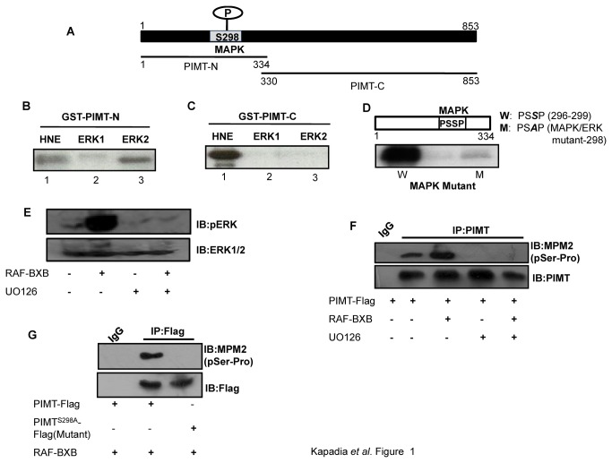
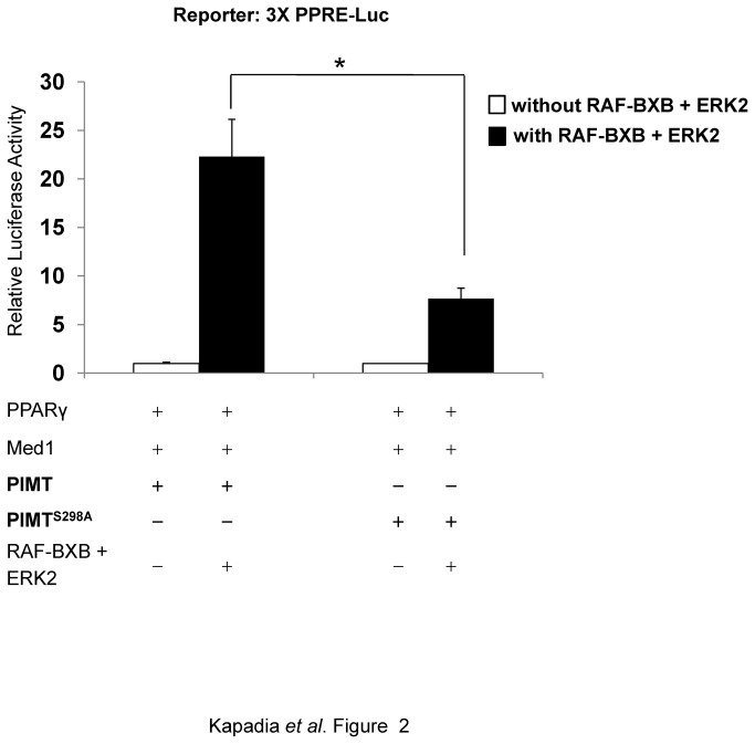
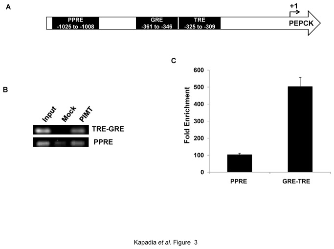
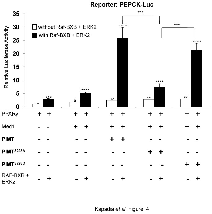
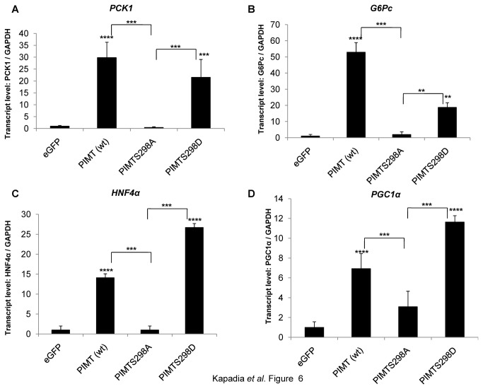
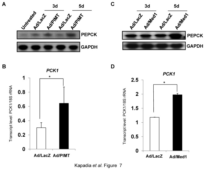
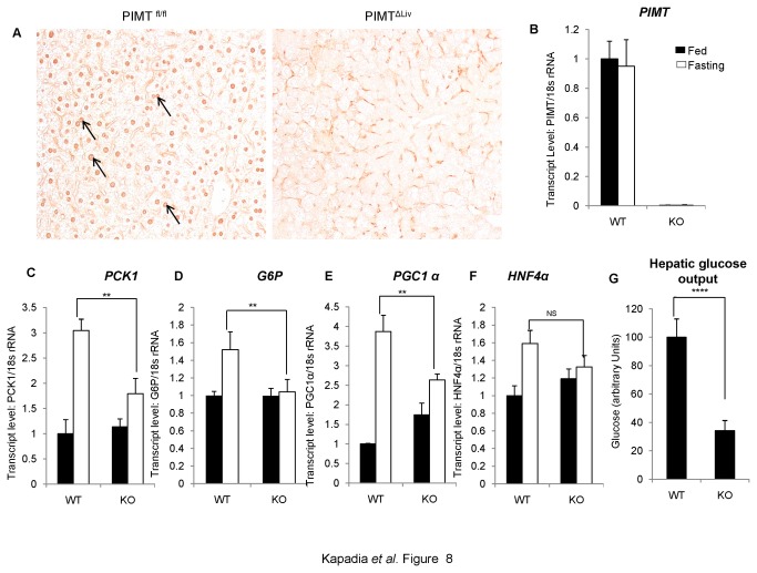
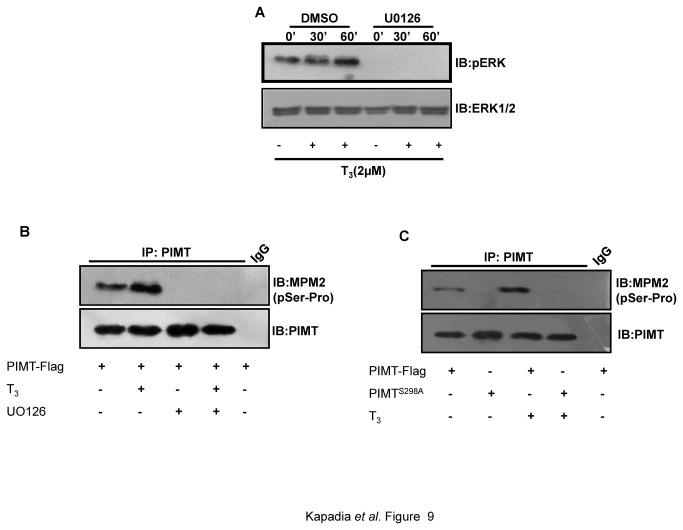
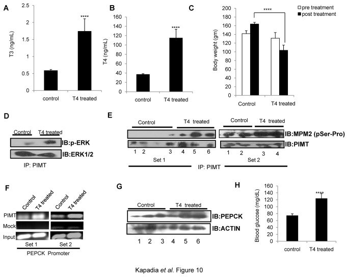
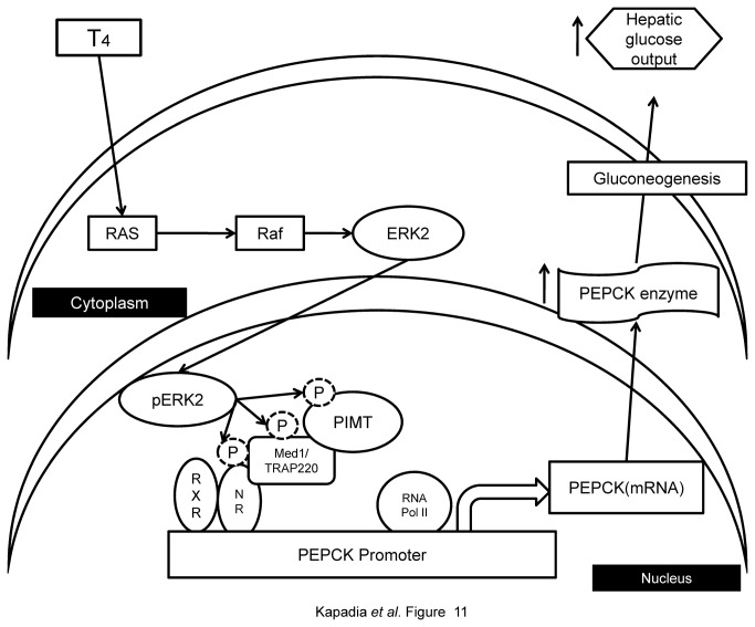
PRIP-Interacting protein with methyl transferase domain (PIMT) serves as a molecular bridge between CREB-binding protein (CBP)/ E1A binding protein p300 (Ep300) -anchored histone acetyl transferase and the Mediator complex sub-unit1 (Med1) and modulates nuclear receptor transcription. Here, we report that ERK2 phosphorylates PIMT at Ser(298) and enhances its ability to activate PEPCK promoter. We observed that PIMT is recruited to PEPCK promoter and adenoviral-mediated over-expression of PIMT in rat primary hepatocytes up-regulated expression of gluconeogenic genes including PEPCK. Reporter experiments with phosphomimetic PIMT mutant (PIMT(S298D)) suggested that conformational change may play an important role in PIMT-dependent PEPCK promoter activity. Overexpression of PIMT and Med1 together augmented hepatic glucose output in an additive manner. Importantly, expression of gluconeogenic genes and hepatic glucose output were suppressed in isolated liver specific PIMT knockout mouse hepatocytes. Furthermore, consistent with reporter experiments, PIMT(S298D) but not PIMT(S298A) augmented hepatic glucose output via up-regulating the expression of gluconeogenic genes. Pharmacological blockade of MAPK/ERK pathway using U0126, abolished PIMT/Med1-dependent gluconeogenic program leading to reduced hepatic glucose output. Further, systemic administration of T4 hormone to rats activated ERK1/2 resulting in enhanced PIMT ser(298) phosphorylation. Phosphorylation of PIMT led to its increased binding to the PEPCK promoter, increased PEPCK expression and induction of gluconeogenesis in liver. Thus, ERK2-mediated phosphorylation of PIMT at Ser(298) is essential in hepatic gluconeogenesis, demonstrating an important role of PIMT in the pathogenesis of hyperglycemia.


Conflict of interest statement

Competing Interests: The authors have declared that no competing interests exist.

Figures

Figure 1
Figure 1. PIMT is a substrate of MAPK.
(A) Schematic diagram of PIMT with a potential phosphorylation site of ERK1/2 at Ser298. PIMT protein was fragmented into 2 parts, PIMT-N (1-334) and PIMT-C (330-853) and fused to GST. (B&C) GST-PIMT-N (B) and GST-PIMT-C (C) bound to glutathione sepharose beads were subjected to kinase reaction in the presence of HeLa nuclear extract [HNE] and constitutively active purified MAPKs ERK1 and ERK2. (D) Glutathione sepharose beads bound GST-PIMT-N [W] and GST-PIMTS298A [M] were subjected to kinase assay with active and purified ERK2. Mutation at MAPK recognition site [PSSP] abolished phosphorylation of PIMT.(E)293T cells were transfected with vector or RAF-BXB and 24 h post transfection cells were treated with DMSO or 10 µM UO126 for 30 min. Subsequently cells were lysed and resolved on 10% SDS-PAGE and probed with Anti-pERK1/2. Blots were stripped and reprobed withAnti-ERK1/2. (F & G) 293T cells were transfected with pCMV-PIMT Flag (F, G) or pCMV- PIMTS298A Flag (G) along with RAF-BXB (F, G) and cells were treated with UO126 where indicated (F). Post transfection cells were cultured in DMEM containing 1% FBS overnight, PIMT was immunoprecipitated with Anti-PIMT followed by separation on 10% SDS-PAGE and probed with Anti-MPM2 (F, G). Blots were stripped and reprobed with Anti-PIMT (F) or Anti-Flag (G) as mentioned.
Figure 2
Figure 2. Phosphorylation of PIMT by RAF-BXB/ERK2 potentiates Med1 dependent transcriptional activity.
HeLa cells were transiently cotransfected with 3X-PPRE driven luciferase construct and PPARγ, Med1 and PIMT or PIMTS298A encoding constructs along with or without RAF-BXB and ERK2. Thirty hours post transfection, cells were lysed and luciferase activity was measured. The values were normalized with corresponding β-galactosidase activity and expressed relative to PIMT (column 1) which was set to 1. Data are representative of 5 independent experiments. Statistical analysis was performed using Student’s t-test (unpaired, two-tailed).* p<0.05.
Figure 3
Figure 3. PIMT is recruited to PEPCK promoter.
(A) A schematic diagram of PEPCK promoter showing PPRE, GRE and TRE. (B&C) Chromatin-immunoprecipitation assay was performed in HepG2 using Anti-PIMT or non specific Anti-Rabbit IgG on human PEPCK promoter (B)and quantified using ChIP qPCR(C).
Figure 4
Figure 4. ERK2 dependent phosphorylation of PIMT increases PEPCK promoter activity.
293T were transfected with pGL3-PEPCK promoter and other constructs as mentioned in Figure 2 legend. Thirty hours post transfection, cells were lysed and luciferase activity was measured. The values were normalized with corresponding Renilla luciferase activity and expressed relative to PPARγ (unphosphorylated) (column 1) which was set to 1. Data are representative of 3 independent experiments. Statistical analysis was performed using one way ANOVA followed by Bonferroeni’s post hoc test. *p<0.001.
Figure 5
Figure 5. Phosphorylation of PIMT at Ser298 is essential for its ability to augment hepatic glucose output.
Primary rat hepatocytes were infected with Ad/PIMT or Ad/PIMTS298A or Ad/PIMTS298D and/or Ad/Med1. Ad/eGFP infection was used as control. After 24 h of infection, the cells were cultured in glucose production medium for 6 h and amount of glucose released in cell supernatants was measured. The values were normalized to corresponding total protein content and expressed relative to Ad/eGFP control which was considered as 1. The inset shows the result of glucose output data in Ad/PIMT or Ad/PIMTS298A or Ad/PIMTS298D infected primary hepatocytes. Data are a representative of 3 independent experiments. Statistical analysis was performed using one way ANOVA followed Bonferroeni’s post-hoc test. ** p<0.01, *** p<0.005, **** p<0.0001 vs Ad/eGFP.
Figure 6
Figure 6. Phosphorylation of PIMT at Ser298 is required for its ability to enhance gluconeogenic genes.
Primary rat hepatocytes were infected with Ad/eGFP (control) or Ad/PIMT or Ad/PIMTS298A or Ad/PIMTS298D and/or Ad/Med1. After 24 h of infection, total RNA was isolated and the expression of (A) PCK1, (B)G6Pc(C)HNF4α and (D)PGC1α was determined using qPCR. The values were normalized to corresponding GAPDH expression and expressed relative to Ad/eGFP control which was considered as 1. Data are a representative of 3 independent experiments. Statistical analysis was performed using one way ANOVA followed Bonferroeni’s post-hoc test. ** p<0.01, *** p<0.005, **** p<0.0001 vs Ad/eGFP.
Figure 7
Figure 7. Overexpression of PIMT or Med1 increases hepatic PEPCK expression in mice.
Mice were injected via tail vein with Ad/LacZ or Ad/PIMT (A & B) or Ad/Med1 (C & D). After 5 days of injection mice were sacrificed and total RNA was isolated from liver. The expression of PEPCK was determined by northern blotting (A and C) or by qPCR (B & D). Blots were re-probed for GAPDH to ensure equal loading (A & C).
Figure 8
Figure 8. Ablation of PIMT expression in liver reduces gluconeogenesis
(A) De-paraffinized 5 μm thin sections were immunohistochemically stained using PIMT antibody in WT and KO mice of liver tissue. (B) Total RNA was isolated from liver tissue and qPCR analysis of PIMT in PIMTfl/fl (WT) and PIMTΔLiv (KO) livers was performed. (C-F) WT and KO mice were fasted for 72h. RNA was extracted and qPCR was performed for PCK1 (C), G6P (D), PGC1α (E) and HNF4α (F). (G) Primary hepatocytes were isolated from WT and KO mice. Post 24h of isolation, cells were culture in glucose production medium for 6 h. Amount of glucose released was estimated and the values were normalized to the corresponding protein content. The values are expressed relative to the normalized WT control group. Data are representative of three independent experiments. Statistical analysis was performed using unpaired Student’s t-test **p<0.01, ****p<0.001, NS: Non-Significant.
Figure 9
Figure 9. Thyroid hormone induced ERK activation is required for phosphorylation of PIMT at Ser298.
(A) 293T cells were stimulated with T3 (2µM) or vehicle control DMSO for the indicated time points and the phosphorylation of pERK1/2 was examined by western blotting. Blots were re-probed with ERK1/2 antibody to ensure equal loading. (B-C) 293T cells were transfected with pCMV-PIMT Flag (B, C) or pCMV- PIMTS298A Flag (C) and cells were treated with or without T3 (2µM) (B,C) and UO126 where indicated (B). Post transfection cells were cultured in DMEM containing 1% FBS overnight followed by separation on 10% SDS-PAGE and probed with Anti-MPM2 (B, C). Blots were stripped and reprobed with Anti-PIMT (B, C).
Figure 10
Figure 10. Thyroid hormone mediated ERK activation stimulates PIMT phosphorylation and enhances hepatic gluconeogenesis in rats.
Rats (n=5) were injected intraperitoneally (0.1mL/animal) with L-thyroxine solution for 2 weeks. Post treatment animals were sacrificed, serum was collected for detecting levels of T3 (A) and T4 (B) and whole body weight (C) were measured. (D) PIMT immunoprecipitated lysates from liver tissue were analyzed by western blotting with phospho-ERK1/2 and total ERK1/2 antibodies. (E)PIMT was immunoprecipitated from liver tissue lysates (Set1: 3 control and 3 treated; Set2: 2 control and 2 treated) separated on 10% SDS PAGE and probed with Anti-MPM2 followed by Anti-PIMT as mentioned. (F) Chromatin immunoprecipitation assay was performed in liver tissue lysates (one control and one treated in both Set1 and Set2) using Anti-PIMT or mock Anti-goat IgG on rat PEPCK promoter. (G) The liver homogenates were analyzed using Anti-PEPCK (top panel) or Anti beta actin (bottom panel). (H) Overnight fasting blood sugar was measured using glucose strip.
Figure 11
Figure 11. Schematicrepresentation of MAPK/ERK mediated PIMT-dependent regulation of hepatic gluconeogenesis.
In response to MAPK induction by T4, ERK2 phosphorylates PIMT (at Ser298 position) which is in complex with Med1 and nuclear receptor (NR). Phosphorylation of PIMT increases Med1-dependent transcriptional activity of gluconeogenic genes leading to enhanced hepatic gluconeogenesis.

Similar articles

Cited by

References

    1. Zhu Y, Qi C, Cao WQ, Yeldandi AV, Rao MS et al. (2001) Cloning and characterization of PIMT, a protein with a methyltransferase domain, which interacts with and enhances nuclear receptor coactivator PRIP function. Proc Natl Acad Sci U S A 98: 10380-10385. doi:10.1073/pnas.181347498. PubMed: 11517327. - DOI - PMC - PubMed
    1. Franke J, Gehlen J, Ehrenhofer-Murray AE (2008) Hypermethylation of yeast telomerase RNA by the snRNA and snoRNA methyltransferase Tgs1. J Cell Sci 121: 3553-3560. doi:10.1242/jcs.033308. PubMed: 18840651. - DOI - PubMed
    1. Komonyi O, Pápai G, Enunlu I, Muratoglu S, Pankotai T et al. (2005) DTL, the Drosophila homolog of PIMT/Tgs1 nuclear receptor coactivator-interacting protein/RNA methyltransferase, has an essential role in development. J Biol Chem 280: 12397-12404. PubMed: 15684427. - PubMed
    1. Jia Y, Viswakarma N, Crawford SE, Sarkar J, Sambasiva Rao M et al. (2012) Early embryonic lethality of mice with disrupted transcription cofactor PIMT/NCOA6IP/Tgs1 gene. Mech Dev 129: 193-207. doi:10.1016/j.mod.2012.08.002. PubMed: 22982455. - DOI - PMC - PubMed
    1. Kornberg RD (2007) The molecular basis of eukaryotic transcription. Proc Natl Acad Sci U S A 104: 12955-12961. doi:10.1073/pnas.0704138104. PubMed: 17670940. - DOI - PMC - PubMed

Publication types

MeSH terms

Substances