. 2013 Dec 17;8(12):e84553.
doi: 10.1371/journal.pone.0084553. eCollection 2013.

Intestinal intraepithelial lymphocyte-enterocyte crosstalk regulates production of bactericidal angiogenin 4 by Paneth cells upon microbial challenge

Affiliations

Intestinal intraepithelial lymphocyte-enterocyte crosstalk regulates production of bactericidal angiogenin 4 by Paneth cells upon microbial challenge

Catherine R Walker et al. PLoS One. .

Abstract

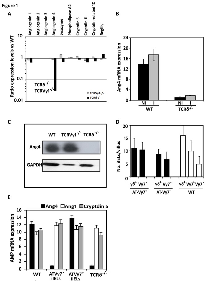
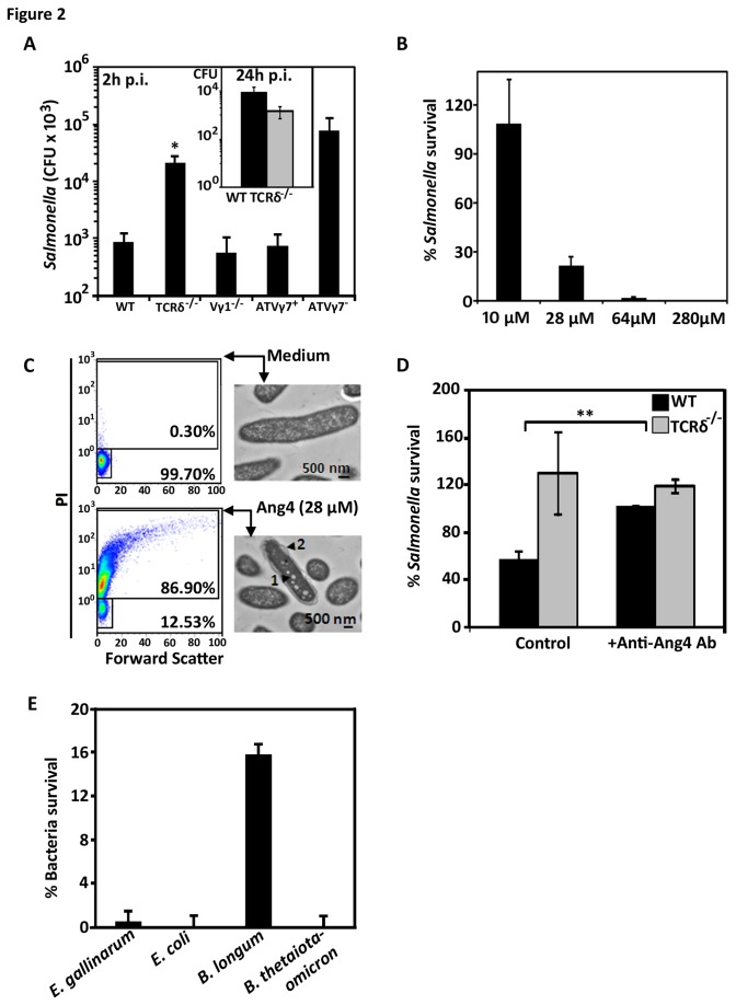
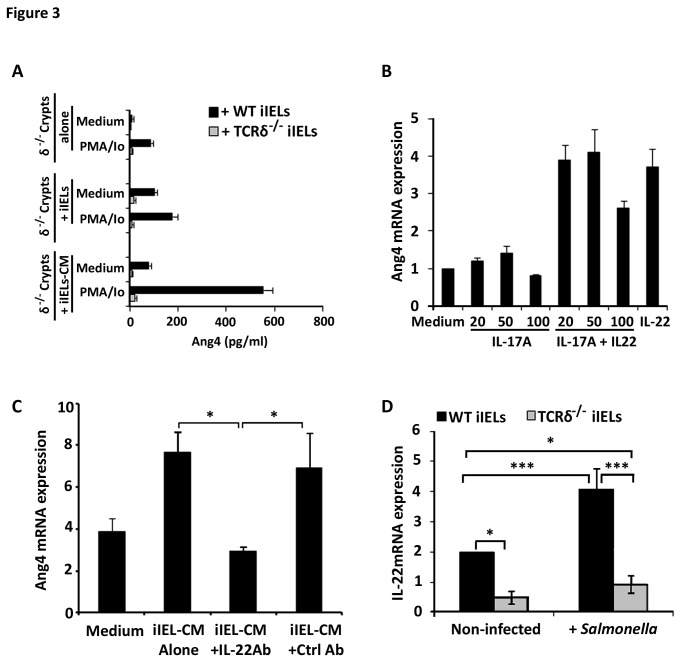
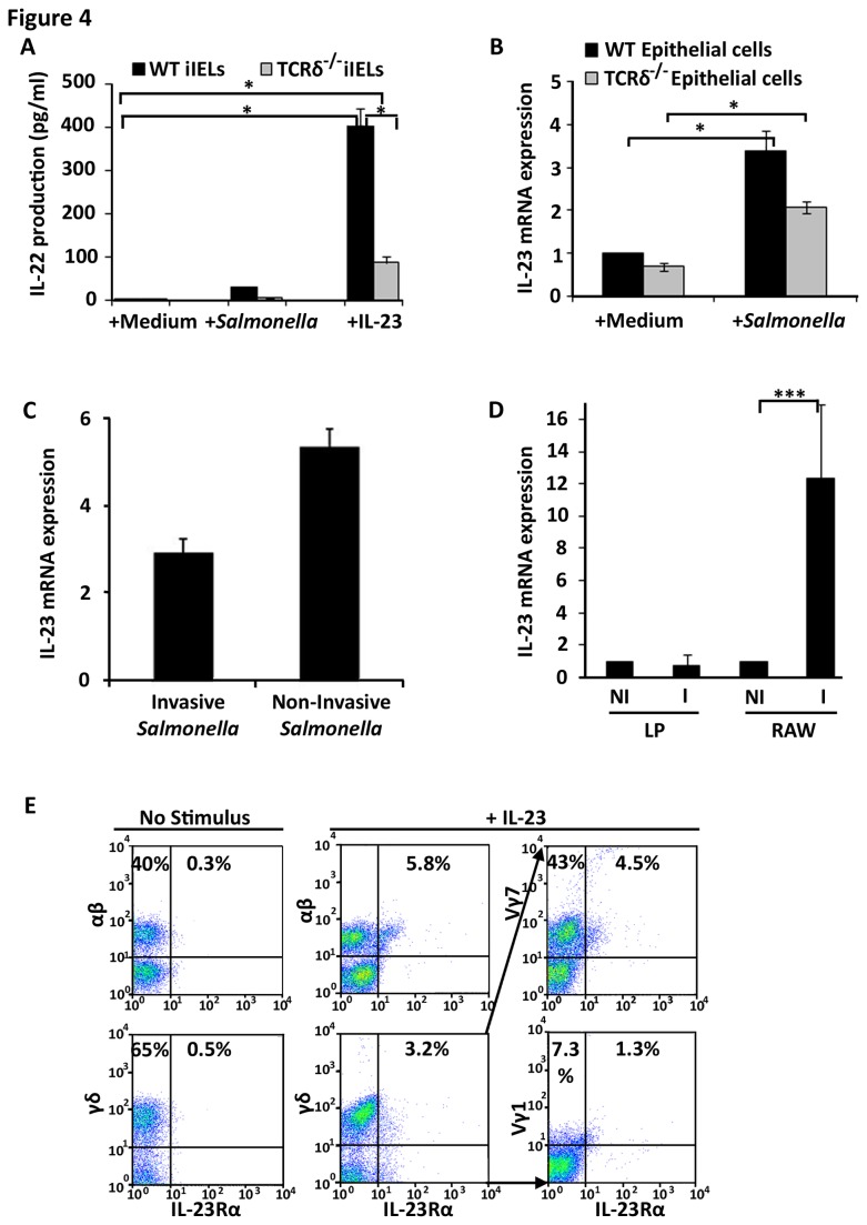
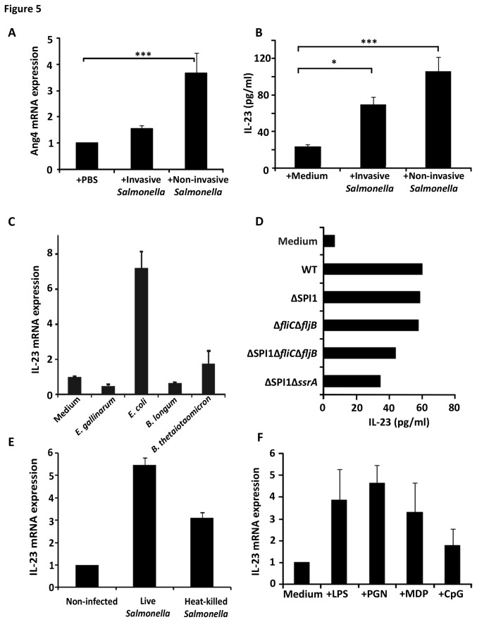
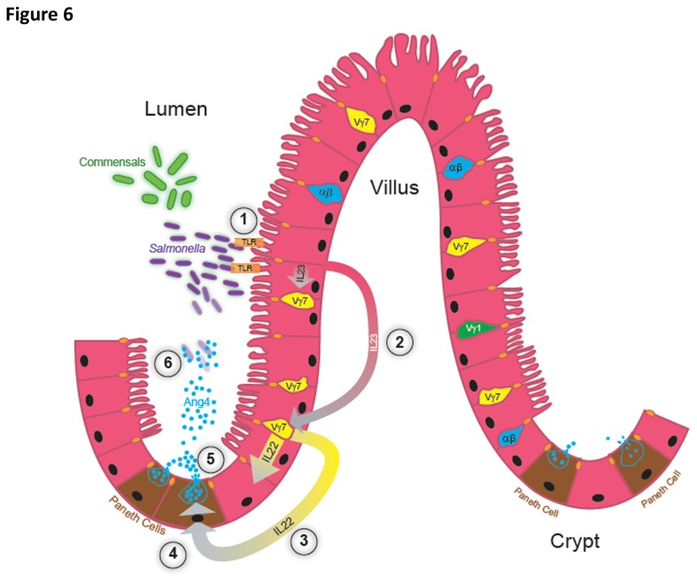
Antimicrobial proteins influence intestinal microbial ecology and limit proliferation of pathogens, yet the regulation of their expression has only been partially elucidated. Here, we have identified a putative pathway involving epithelial cells and intestinal intraepithelial lymphocytes (iIELs) that leads to antimicrobial protein (AMP) production by Paneth cells. Mice lacking γδ iIELs (TCRδ(-/-)) express significantly reduced levels of the AMP angiogenin 4 (Ang4). These mice were also unable to up-regulate Ang4 production following oral challenge by Salmonella, leading to higher levels of mucosal invasion compared to their wild type counterparts during the first 2 hours post-challenge. The transfer of γδ iIELs from wild type (WT) mice to TCRδ(-/-) mice restored Ang4 production and Salmonella invasion levels were reduced to those obtained in WT mice. The ability to restore Ang4 production in TCRδ(-/-) mice was shown to be restricted to γδ iIELs expressing Vγ7-encoded TCRs. Using a novel intestinal crypt co-culture system we identified a putative pathway of Ang4 production initiated by exposure to Salmonella, intestinal commensals or microbial antigens that induced intestinal epithelial cells to produce cytokines including IL‑23 in a TLR-mediated manner. Exposure of TCR-Vγ7(+) γδ iIELs to IL-23 promoted IL‑22 production, which triggered Paneth cells to secrete Ang4. These findings identify a novel role for γδ iIELs in mucosal defence through sensing immediate epithelial cell cytokine responses and influencing AMP production. This in turn can contribute to the maintenance of intestinal microbial homeostasis and epithelial barrier function, and limit pathogen invasion.


Conflict of interest statement

Competing Interests: The authors have declared that no competing interests exist.

Figures

Figure 1
Figure 1. γδ T cells modulate the production of Ang4.
(A) Expression levels of Paneth cell AMPs in the intestinal mucosa of naïve wild type (WT), TCRδ-/- and TCRVγ1‑/- mice as determined by RNA microarrays [82]. Levels are displayed as a ratio of the value obtained in TCRδ-/- or TCRVγ1-/- versus WT samples. (B) The level of Ang4 mRNA in the intestinal epithelium of TCRδ-/- and WT mice was determined both prior to (NI) and 2h post-oral challenge and infection (I) with Salmonella Typhimurium by qPCR. Data (mean±SEM) are expressed relative to levels of β‑actin mRNA and were collated from 6 RNA samples in each group. (C) Ang4 protein levels in the intestine of naïve WT, TCRδ‑/- and Vγ1‑/- mice determined by immunoblotting. Membranes were stripped and re-probed with an anti-GAPDH antibody. The results shown are representative of those obtained using 4-6 mice of each strain. (D) Number of γδ+ and TCR-Vγ7+ iIELs in intestinal tissue sections by immunohistochemistry 6 weeks after transfusion of γδ iIEL-deficient mice with TCR-Vγ7+ or TCR-Vγ7- iIELs. The data were collated (mean±SEM) by counting stained cells in at least 30 villi per section on a minimum of 5 sections per tissue from 4-6 mice. (E) Ang4 production is restored in iIEL-reconstituted TCRδ‑/- mice to WT levels after transfusion of Vγ7+ (ATVγ7+) iIELs. Levels of Ang4, Ang1 and cryptidin 5 mRNA were determined by qPCR in samples of small intestine obtained from WT, TCRδ-/- and TCRδ-/- mice 6 weeks post‑reconstitution with Vγ7+ or Vγ7- (ATVγ7-) iIELs and 2h after oral challenge with Salmonella. Data (mean±STD) are expressed relatively to levels of β‑actin mRNA and were collated from RNA samples of 4-6 mice of each group.
Figure 2
Figure 2. Vγ7+ iIELs are involved in resisting Salmonella invasion.
(A) SL1344-Tnlux Salmonella population levels in the intestinal (ileal) mucosa of WT, TCRδ-/- and TCRVγ1-/- mice (n=10-12) and in TCRδ-/- mice reconstituted with either Vγ7+ iIELs (ATVγ7+) or Vγ7- iIELs (ATVγ7-) (n=5-6) 2h after oral challenge, (*p<0.01 comparing TCRδ-/- with WT group). Salmonella CFU were quantified as described in the Materials and Methods. Inset: CFU of Salmonella SL1344wt strain in ileal mucosa 24h post-oral challenge. (B) Survival of 1x10⁵ S. Typhimurium SL1344 after 1h exposure to increasing amounts of recombinant Ang4, expressed as a percentage of population treated with PBS only. Data shown (mean±SEM) are representative of three independent experiments, each performed in triplicates. (C) Viability and cell membrane alteration of Ang4-treated Salmonella as assessed by PI staining and flow cytometry, and by transmission electron microscopy. The proportion of viable and dead bacteria after incubation with Ang4 or PBS is indicated by the % values shown in the quadrants. The TEM images are representative of 200‑300 Salmonella cells observed. The black arrowheads indicate regions of vesicle‑like structures (1) and blebbing of the outer membrane (2). (D) Survival of 1x10⁵ CFU S. Typhimurium SL1344 exposed to freshly collected TCRδ-/- and WT crypt exudates in 10mM iPIPES (PIPES containing 137mM NaCl) in presence or absence of anti-Ang4 neutralising antibody (M20; Santa Cruz) (mean±SEM; **p≤0.005). Survival to Ang4 exposure was measured relative to that in non-treated exudates. (E) Survival of 1x10⁵ Enterococcus gallinarum, Escherichia coli, Bifidobacterium longum and Bacteroides thetaiotaomicron commensal bacteria after 1h exposure to 28μM of recombinant Ang4, expressed as a percentage of population treated with PBS only. Data shown are the mean±SEM of three independent experiments.
Figure 3
Figure 3. Paneth cells produce Ang4 in response to IL‑22 of which γδ iIELs are a source.
(A) ELISA-determined Ang4 protein levels produced by small intestinal crypts (2x10³) from TCRδ-/- mice after culture at 37°C for 4h in media alone (Medium) or in media containing PMA/Io. Additional crypt samples were cultured with 103 WT iIELs (+iIELs) with and without prior in vitro stimulation by PMA/Io or, with conditioned medium (iIEL-CM) obtained from 104 in vitro stimulated WT (black bars) and TCRδ-/- (grey bars) iIELs. Data (mean±SEM) were collated from three experiments. (B) Ang4 mRNA levels (qPCR) detected in isolated intestinal TCRδ-/- crypts/Paneth cells incubated at 37°C for 4h with recombinant murine IL‑17A alone or in combination with recombinant IL‑22 (100ng/ml). Control cultures contained medium or 100ng/ml IL-22 alone. Data (mean±SEM) are expressed relatively to mRNA levels obtained when crypts were exposed to medium alone and were collated from two experiments (see also Figure S2). (C) Anti-IL‑22 antibodies abrogate Ang4 expression by iIEL-CM. Small intestinal crypts from TCRδ-/- mice were cultured at 37°C for 4h with iIEL-CM in the presence or absence of neutralising anti-IL‑22 or control antibodies (Ctrl Ab). Crypt Ang4 mRNA levels were measured by qPCR and expressed relative to β‑actin mRNA, with values (mean±SEM) representative of three experiments. (D) IL‑22 expression is reduced in TCRδ‑/- iIELs. IL‑22 mRNA was isolated from iIELs of WT and TCRδ-/- mice prior to (Non-infected) and 2h post‑challenge with Salmonella by qPCR. The data (mean±SEM) are expressed relative to β‑actin mRNA and were collated from four experiments (*p≤0.05; ***p≤0.001).
Figure 4
Figure 4. iIELs produce IL-22 in response to IL-23, a cytokine produced by intestinal epithelial cells.
(A) iIELs from WT and TCRδ‑/‑ mice were cultured at 37°C for 6h with medium alone or medium containing 1x10⁵ CFU S. Typhimurium or recombinant IL‑23 (100ng/ml) after which supernatants were analysed by IL‑22 ELISA (*p≤0.05). Data (mean±SEM) were collated from three experiments. (B) Intestinal epithelial cells (106) from WT and TCRδ‑/- mice were exposed for 2h to medium alone or medium containing Salmonella at a ratio of 10 bacteria per epithelial cell. IL‑23 mRNA expression was then assessed by qPCR. The data (mean±SEM) were expressed relative to mRNA levels found in WT epithelial cells exposed to medium alone and were collated from three experiments (*p≤0.05). (C) m-ICcl2 intestinal epithelial cells were cultivated on 0.4µm pore size filters and incubated at 37°C for 4h with medium alone, or medium containing invasive or non‑invasive Salmonella after which cells were harvested, RNA extracted and IL‑23 expression analysed by qPCR. Data are expressed relative to mRNA levels obtained on m‑ICc12 cells exposed to medium alone. (D) RNA was purified from lamina propria (LP) cells isolated from orally infected (2h p.i.) and non‑infected WT mice. As a positive control for IL-23 production, cultivated RAW264.7 macrophages (RAW) were analysed pre- (NI) and post-infection (I) (6h p.i.) with Salmonella. Data are shown as mean±STD relative to mRNA levels in non‑infected samples, and are representative of 5 and 4 experiments, respectively. (E) iIELs from WT mice were cultured at 37°C for 2h with medium alone (No Stimulus) or with medium containing IL‑23. The distribution of IL-23Rα expression among Vγ1 and Vγ7 iIELs was determined by flow cytometry from 4-colour/antibody staining protocols: combining CD3, TCR-γδ, IL-23R and either TCR-Vγ7 or TCR-Vγ1 antibodies with a gating strategy of selecting CD3+, TCR-γδ+, IL-23R+ events (middle panels) and then analysing them for Vγ7+, IL-23Rα+ and Vγ7-, IL-23Rα+ events (far right hand panels). The profiles shown are representative of three experiments with the percentage values representing the frequency of positive cells in the designated quadrants.
Figure 5
Figure 5. Requirements for microbe-induced Ang4 and IL‑23 production by intestinal epithelial cells.
(A) Invasive or non-invasive Salmonella strains (4x10⁷ CFU in PBS) were injected into exteriorised intestinal ligated loops of WT mice. Four hours later mucosal RNA was isolated and Ang4 mRNA expression analysed by qPCR. The data (mean±SEM) are expressed relative to mRNA levels obtained from loops exposed to PBS alone (n=3; ***p<0.001). (B) IL‑23 protein levels assessed by ELISA (mean±SEM) from intestinal epithelial cells of WT mice exposed for 12h to medium alone, (+Medium) or media containing invasive or non‑invasive Salmonella strains at a ratio of 10 bacteria per epithelial cell (n=3; ***p<0.001, *p<0.05). (C) m-ICc12 intestinal epithelial cells were cultured for 4h with medium alone (control) or containing live cells of various intestinal commensal bacteria at a ratio of 10 bacteria per epithelial cell. IL‑23 mRNA expression was assessed by qPCR. Data (mean±SEM) were expressed relatively to mRNA levels in control samples (n=4). (D) IL‑23 protein production measured by ELISA in intestinal epithelial cells from WT mice cultured at 37°C for 12h with medium alone or containing either, live WT (SL1344) or various Salmonella mutant strains that are non-invasive (SL1344ΔSPI1), invasive but non-flagellated (JH3220=SL1344ΔfliCΔfljB), non-invasive and non-flagellated (JH3515=SL1344∆SPI1ΔfliCΔfljB) or non-invasive, flagellated but unable to transcytose flagellin (JH3574=SL1344∆SPI1ΔssrA) (n=3; mean±SEM). (E) IL‑23 mRNA expression assessed by qPCR in m-ICc12 epithelial cells cultured at 37°C for 4h with medium alone or containing live or heat‑killed WT Salmonella SL1344 cells at a ratio of 10 bacteria per epithelial cell. Data (n=4; mean±SEM) are expressed relative to mRNA levels obtained in non‑infected cells. (F) IL‑23 mRNA levels analysed by qPCR in intestinal epithelial cells from WT mice cultured at 37°C for 2h with medium alone or containing lipopolysaccharide (LPS), peptidoglycan (PGN), muramyl dipeptide (MDP) or methylated DNA (CpG). Data (n=3; mean±SEM) are expressed relative to mRNA levels obtained in cells exposed to medium alone.
Figure 6
Figure 6. Hypothetical model of the production of Ang4 in response to microbial challenge.
(1) Upon exposure to Salmonella or recognition of commensal bacteria or MAMPs, intestinal epithelial cells secrete IL‑23, in a TLR‑dependent manner in the case of Salmonella. (2) Via extracellular or transcellular routes, epithelial cells secrete IL‑23 (pink to grey gradient arrows) that binds to its cognate receptor IL‑23R expressed by γδ iIELs. (3) IL-23R+ iIELs enriched in Vγ7+ cells respond to IL-23 by secreting IL‑22 (yellow to grey gradient arrows). Via extracellular or transcellular routes IL‑22 acts on IL-22R-bearing Paneth cells up-regulating Ang4 transcription (4) and/or secretion (5) of pre-formed protein stored in intracellular granules. Ang4 is secreted into the lumen at levels sufficient (5) to effectively kill Salmonella located in the vicinity of the intestinal tissue (6), helping to protect it from proliferation of and invasion by the pathogen.

Similar articles

Cited by

References

    1. Sansonetti PJ (2004) War and peace at mucosal surfaces. Nat Rev Immunol 4: 953-964. doi:10.1038/nri1499. PubMed: 15573130. - DOI - PubMed
    1. Ayabe T, Satchell DP, Wilson CL, Parks WC, Selsted ME et al. (2000) Secretion of microbicidal α-defensins by intestinal Paneth cells in response to bacteria. Nat Immunol 1: 113-118. doi:10.1038/77783. PubMed: 11248802. - DOI - PubMed
    1. Salzman NH, Ghosh D, Huttner KM, Paterson Y, Bevins CL (2003) Protection against enteric salmonellosis in transgenic mice expressing a human intestinal defensin. Nature 422: 522-526. doi:10.1038/nature01520. PubMed: 12660734. - DOI - PubMed
    1. Wilson CL, Ouellete AJ, Satchell DP, Ayabe T, Lopez-Boado YS et al. (1999) Regulation of intestinal α-defensin activation by the metalloproteinase matrilysin in innate host defence. Science 286. - PubMed
    1. Salzman NH, Hung K, Haribhai D, Chu H, Karlsson-Sjöberg J et al. (2010) Enteric defensins are essential regulators of intestinal microbial ecology. Nat Immunol 11: 76-82. doi:10.1038/ni.1825. PubMed: 19855381. - DOI - PMC - PubMed

Publication types

MeSH terms