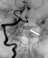
Marginal sinus fistula supplied exclusively by vertebral artery feeders
- PMID: 24358414
- PMCID: PMC3868244
Marginal sinus fistula supplied exclusively by vertebral artery feeders
Abstract
A 54-year-old woman is reported with severe pulsatile tinnitus. Digital subtraction angiography demonstrated dural arteriovenous fistula of the marginal sinus with feeders arising exclusively from bilateral vertebral arteries. Patient underwent successful transarterial Onyx embolization with complete angiographic and clinical cure.
Keywords: Onyx; arteriovenous malformation; dural fistula; marginal sinus; vertebral artery.
Figures


Similar articles
-
Safety of Onyx Transarterial Embolization of Skull Base Dural Arteriovenous Fistulas from Meningeal Branches of the External Carotids also Fed by Meningeal Branches of Internal Carotid or Vertebral Arteries.Clin Neuroradiol. 2018 Dec;28(4):579-584. doi: 10.1007/s00062-017-0615-7. Epub 2017 Aug 11. Clin Neuroradiol. 2018. PMID: 28801711
-
Middle meningeal artery: Gateway for effective transarterial Onyx embolization of dural arteriovenous fistulas.Clin Anat. 2016 Sep;29(6):718-28. doi: 10.1002/ca.22733. Epub 2016 Jun 7. Clin Anat. 2016. PMID: 27148680
-
An Onyx tunnel: reconstructive transvenous balloon-assisted Onyx embolization for dural arteriovenous fistula of the transverse-sigmoid sinus.J Neurosurg. 2018 Oct;129(4):922-927. doi: 10.3171/2017.5.JNS17287. Epub 2017 Nov 17. J Neurosurg. 2018. PMID: 29148903
-
[Dural arteriovenous fistula of the transverse sigmoid sinus after transvenous embolization of the carotid cavernous fistula].No To Shinkei. 1999 Dec;51(12):1065-9. No To Shinkei. 1999. PMID: 10654304 Review. Japanese.
-
Complete Obliteration of a Foramen Magnum Dural Arteriovenous Fistula by Microsurgery After Failed Endovascular Treatment Using Onyx: Case Report and Literature Review.World Neurosurg. 2020 Dec;144:43-49. doi: 10.1016/j.wneu.2020.08.077. Epub 2020 Aug 15. World Neurosurg. 2020. PMID: 32805464 Review.
Cited by
-
Dural arteriovenous fistula of the lateral foramen magnum region: A review.Interv Neuroradiol. 2018 Aug;24(4):425-434. doi: 10.1177/1591019918770768. Epub 2018 May 4. Interv Neuroradiol. 2018. PMID: 29726736 Free PMC article. Review.
-
Safety of Onyx Transarterial Embolization of Skull Base Dural Arteriovenous Fistulas from Meningeal Branches of the External Carotids also Fed by Meningeal Branches of Internal Carotid or Vertebral Arteries.Clin Neuroradiol. 2018 Dec;28(4):579-584. doi: 10.1007/s00062-017-0615-7. Epub 2017 Aug 11. Clin Neuroradiol. 2018. PMID: 28801711
References
-
- Tamargo RJ, Huang J. Brain arteriovenous malformations (AVMs) and dural arteriovenous fistulas (DAVFs): rare and formidable lesions. Neurosurg Clin N Am. 2012;23(1):xiii–xiv. - PubMed
-
- Söderman M, Pavic L, Edner G, Holmin S, Andersson T. Natural history of dural arteriovenous shunts. Stroke. 2008;39(6):1735–9. - PubMed
-
- Al-Shahi R, Bhattacharya JJ, Currie DG, et al. Prospective, population-based detection of intracranial vascular malformations in adults: the Scottish Intracranial Vascular Malformation Study (SIVMS) Stroke. 2003;34(5):1163–9. - PubMed
-
- Brown RD, Jr, Wiebers DO, Torner JC, et al. Incidence and prevalence of intracranial vascular malformations in Olmsted County, Minnesota, 1965 to 1992. Neurology. 1996;46:949–52. - PubMed
LinkOut - more resources
Full Text Sources
