. 2013 Sep 30;3(5):1000158.

Oxidative Lung Damage Resulting from Repeated Exposure to Radiation and Hyperoxia Associated with Space Exploration

Affiliations

Oxidative Lung Damage Resulting from Repeated Exposure to Radiation and Hyperoxia Associated with Space Exploration

Ralph A Pietrofesa et al. J Pulm Respir Med. .

Abstract

Background: Spaceflight missions may require crewmembers to conduct Extravehicular Activities (EVA) for repair, maintenance or scientific purposes. Pre-breathe protocols in preparation for an EVA entail 100% hyperoxia exposure that may last for a few hours (5-8 hours), and may be repeated 2-3 times weekly. Each EVA is associated with additional challenges such as low levels of total body cosmic/galactic radiation exposure that may present a threat to crewmember health and therefore, pose a threat to the success of the mission. We have developed a murine model of combined, hyperoxia and radiation exposure (double-hit) in the context of evaluating countermeasures to oxidative lung damage associated with space flight. In the current study, our objective was to characterize the early and chronic effects of repeated single and double-hit challenge on lung tissue using a novel murine model of repeated exposure to low-level total body radiation and hyperoxia. This is the first study of its kind evaluating lung damage relevant to space exploration in a rodent model.

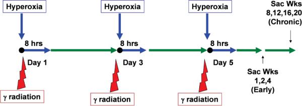
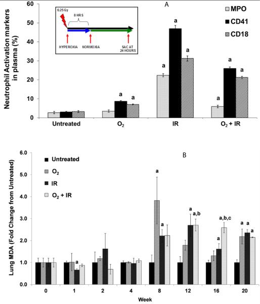
Methods: Mouse cohorts (n=5-15/group) were exposed to repeated: a) normoxia; b) >95% O2 (O2); c) 0.25Gy single fraction gamma radiation (IR); or d) a combination of O2 and IR (O2+IR) given 3 times per week for 4 weeks. Lungs were evaluated for oxidative damage, active TGFβ1 levels, cell apoptosis, inflammation, injury, and fibrosis at 1, 2, 4, 8, 12, 16, and 20 weeks post-initiation of exposure.

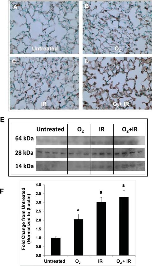
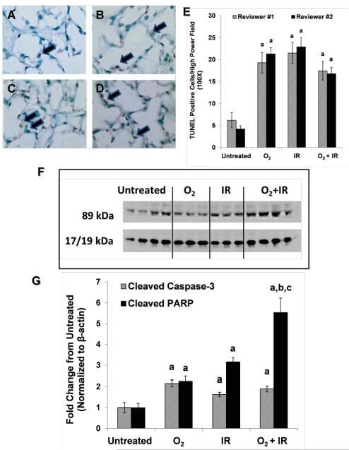
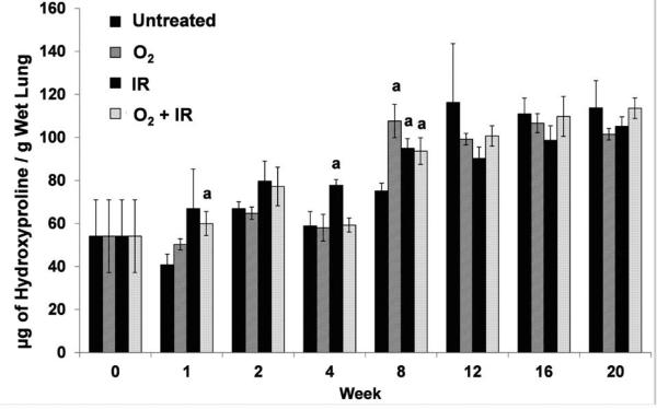
Results: Mouse cohorts exposed to all challenge conditions displayed decreased bodyweight compared to untreated controls at 4 and 8 weeks post-challenge initiation. Chronic oxidative lung damage to lipids (malondialdehyde levels), DNA (TUNEL, cleaved Caspase 3, cleaved PARP positivity) leading to apoptotic cell death and to proteins (nitrotyrosine levels) was elevated all treatment groups. Importantly, significant systemic oxidative stress was also noted at the late phase in mouse plasma, BAL fluid, and urine. Importantly, however, late oxidative damage across all parameters that we measured was significantly higher than controls in all cohorts but was exacerbated by the combined exposure to O2 and IR. Additionally, impaired levels of arterial blood oxygenation were noted in all exposure cohorts. Significant but transient elevation of lung tissue fibrosis (p<0.05), determined by lung hydroxyproline content, was detected as early as 2 week in mice exposed to challenge conditions and persisted for 4-8 weeks only. Interestingly, active TGFβ1 levels in +BAL fluid was also transiently elevated during the exposure time only (1-4 weeks). Inflammation and lung edema/lung injury was also significantly elevated in all groups at both early and late time points, especially the double-hit group.

Conclusion: We have characterized significant, early and chronic lung changes consistent with oxidative tissue damage in our murine model of repeated radiation and hyperoxia exposure relevant to space travel. Lung tissue changes, detectable several months after the original exposure, include significant oxidative lung damage (lipid peroxidation, DNA damage and protein nitrosative stress) and increased pulmonary fibrosis. These findings, along with increased oxidative stress in diverse body fluids and the observed decreases in blood oxygenation levels in all challenge conditions (whether single or in combination), lead us to conclude that in our model of repeated exposure to oxidative stressors, chronic tissue changes are detected that persist even months after the exposure to the stressor has ended. This data will provide useful information in the design of countermeasures to tissue oxidative damage associated with space exploration.

Keywords: Apoptosis; Bronchoalveolar lavage; Caspase 3; Double-hit; Extravehicular activity; Hyperoxia; Inflammation; Lung fibrosis; Lung injury; Mouse model; Nitrotyrosine; Oxidative stress; PARP; Radiation pneumonopathy; Space exploration; TGF-β1; TUNEL; Total body irradiation.


Figures

Figure 1
Figure 1. Experimental Plan of In Vivo Animal Exposure
Mouse cohorts (n=5-15 / group) were exposed to a double-hit challenge of 0.25 Gy total body ionizing gamma irradiation (IR) and 100% pO2 for 8 hours followed by intermittent normoxia (21% pO2). This cycle was performed 3 times a week for 1, 2, and 4 weeks. Mice were sacrificed at weeks 1, 2, and 4 weeks (early) and 8, 12, 16, and 20 weeks (chronic). A total of 24 hours of hyperoxia / radiation exposure weekly simulates the maximum allowed weekly extravehicular activity (EVA) exposure for crewmembers on lengthy spaceflights.
Figure 2
Figure 2. Kinetic study of the effect of hyperoxia, radiation, or double-hit combination on blood oxygenation
Separate cohorts of mice (n=5-15 / group) were exposed to 100% O2 for 8 hours only (O2), 0.25 Gy total body ionizing gamma irradiation (IR) only, or a double-hit combination of both challenges (O2+IR) followed by intermittent normoxia (21% pO2) for repeated cycles 3 times a week for 4 weeks. Mice were sacrificed at weeks 1, 2, and 4 weeks (acute) and 8, 12,,16, and 20 weeks (chronic). Arterial blood oxygenation was determined prior to sacrifice 8, 12, 16, and 20 weeks. Data is represented as mean ± SEM. Letter a indicates p< 0.05 for all challenges vs. untreated at each respective time point. Letter b indicates p< 0.05 for O2+IR vs. O2 at each respective time point. Letter c indicates p< 0.05 for O2+IR vs IR at each respective time point.
Figure 3
Figure 3. Evaluation of the effect of hyperoxia, radiation, or double-hit combination on lung inflammation and damage/edema
(Panel A) Separate cohorts of mice (n=5-15 / group) were exposed to 100% O2 for 8 hours only (O2), 0.25 Gy total body ionizing gamma irradiation (IR) only, or a double-hit combination of both challenges (O2+IR) followed by intermittent normoxia (21% pO2) for repeated cycles 3 times a week for 4 weeks. Mice were sacrificed at weeks 1, 2, and 4 weeks (acute) and 8, 12, 16 and 20 weeks (chronic). BAL white blood cell counts were evaluated as a measure of lung injury. (Panel B) BAL was performed at the time of sacrifice and BAL proteins determined as a measure of lung damage. Data is represented as mean fold change from untreated at each respective time point ± SEM. Letter a indicates p< 0.05 for all challenges vs. untreated at each respective time point. Letter b indicates p< 0.05 for O2+IR vs. O2 at each respective time point. Letter c indicates p< 0.05 for O2+IR vs IR at each respective time point.
Figure 4
Figure 4. Kinetics of inflammatory cell activation and oxidative damage in mouse lungs after repeated cycles of O2, IR or O2+IR exposure
(Panel A) Mice were exposed to 100% O2 for 8 hours only (O2), 0.25 Gy total body ionizing gamma irradiation (IR) only, or a double-hit combination of both challenges (O2+IR) followed by 16 hours of normoxia (21% pO2) for a single cycle. Whole blood was collected and leukocyte analysis of MPO, CD41, and CD18 was performed using flow cytometry. (Panel B) Separate cohorts of mice (n=5-15 / group) were exposed to 100% O2 for 8 hours only (O2), 0.25 Gy total body ionizing gamma irradiation (IR) only, or a double-hit combination of both challenges (O2+IR) followed by intermittent normoxia (21% pO2) for repeated cycles 3 times a week for 4 weeks. Mice were sacrificed at weeks 1, 2, and 4 weeks (early) and 8, 12, 16, and 20 weeks (chronic). Oxidative modification of lung tissues was measured by determining MDA levels in lung. Data is represented as mean fold change from untreated at each respective time point ± SEM. Letter a indicates p< 0.05 for all challenges vs. untreated at each respective time point. Letter b indicates p< 0.05 for O2+IR vs. O2 at each respective time point. Letter c indicates p< 0.05 for O2+IR vs IR at each respective time point.
Figure 5
Figure 5
Determination of oxidative stress in mouse plasma, BAL fluid, and urine at week 20 post-initiation of repeated cycles of O2, IR or O2+IR exposure Separate cohorts of mice (n=5-15 / group) were exposed to 100% O2 for 8 hours only (O2), 0.25 Gy total body ionizing gamma irradiation (IR) only, or a double-hit combination of both challenges (O2+IR) followed by intermittent normoxia (21% pO2) for repeated cycles 3 times a week for 4 weeks. At week 20, mouse plasma, BAL fluid, and urine were collected. Oxidative modification was determined by measuring MDA levels. Data is represented as mean ± SEM. Letter a indicates p< 0.05 for each challenge condition cohort vs. untreated.
Figure 6
Figure 6
Effects of repeated O2, IR and combined challenge on Nitrotyrosine formation, during the late phase post termination of the challenge exposure. Separate cohorts of mice were exposed to 100% O2 for 8 hours only (O2), 0.25 Gy total body ionizing gamma irradiation (IR) only, or a double-hit combination of both challenges (O2+IR) followed by intermittent normoxia (21% pO2) for repeated cycles 3 times a week for 4 weeks. Mice were sacrificed at 20 weeks (chronic). Panels A-D Representative sections from paraffin lung sections stained for nitrotyrosine (brown staining) from untreated (A), O2 (B), IR (C) and O2+IR (D) animals. Sections were counterstained with crystal blue. Panel E Representative western blot validation of NT formation in lung homogenates from the same mouse cohorts as above. Panel F Densitometric analysis with beta actin normalization of the combined levels of all 3 major NT bands for each cohort. Data is represented as mean fold change from untreated at each respective time point ± SEM. Letter a indicates p< 0.05 for all challenges vs. untreated at each respective time point.
Figure 7
Figure 7. Determination of oxidative DNA damage using TUNEL staining on lung sections at various times post-initiation of repeated cycles of O2, IR or O2+IR exposure
Panels A-D Histological lung sections of representative animals from each cohort (O2, IR, O2+IR) processed for TUNEL positivity (late phase, 20 weeks). Negative controls (performed simultaneously but without TdT) were examined at the same time. Panel E Cells were counted as % positive cells per high power field (400X) in 10 fields per condition. Data is represented as mean ± SEM. p< 0.05 for each challenge condition cohort vs. untreated as determined by two independent reviewers. Panel F Western blot for cleaved caspase 3 and cleaved PARP. Panel G Densitometric analysis of representative blot. Data is represented as mean fold change from untreated at each respective time point ± SEM. Letter a indicates p< 0.05 for all challenges vs. untreated at each respective time point. Letter b indicates p< 0.05 for O2+IR vs. O2 at each respective time point. Letter c indicates p< 0.05 for O2+IR vs IR at each respective time point.
Figure 8
Figure 8. Kinetics of pulmonary fibrotic changes in mouse lungs after repeated cycles of O2, IR or O2+IR exposure
Separate cohorts of mice (n=5-15 / group) were exposed to 100% O2 for 8 hours only (O2), 0.25 Gy total body ionizing gamma irradiation (IR) only, or a double-hit combination of both challenges (O2+IR) followed by intermittent normoxia (21% pO2) for repeated cycles 3 times a week for 4 weeks. Mice were sacrificed at weeks 1, 2, and 4 weeks (acute) and 8, 12, 16 and 20 weeks (chronic). Lung fibrosis was determined by measuring lung hydroxyproline content. Data is represented mean ± SEM. Letter a indicates p< 0.05 each challenge cohort as it compares with the untreated control at the respective time point.
Figure 9
Figure 9. Determination of Active TGF-β1 levels in mouse BAL fluid at various times post-initiation of repeated cycles of O2, IR or O2+IR exposure
Separate cohorts of mice (n=5-15 / group) were exposed to 100% O2 for 8 hours only (O2), 0.25 Gy total body ionizing gamma irradiation (IR) only, or a double-hit combination of both challenges (O2+IR) followed by intermittent normoxia (21% pO2) for repeated cycles 3 times a week for 4 weeks. At 1,2,4,8,12,16 and 20 weeks post exposure, BAL fluid were collected. Active TGF-β1 levels were determined by ELISA. Data is represented as mean ± SEM. Letter a indicates p< 0.05 for each challenge condition cohort vs. untreated.
Figure 10
Figure 10
Mechanism of damage in mouse lungs after repeated cycles of O2, IR or O2+IR exposure.

Similar articles

Cited by

References

    1. Holloway RJ, Leong GF, Ainsworth EJ, Albright ML, Baum SJ. Recovery from radiation injury in the hamster as evaluated by the split-dose technique. USNRDL-TR-1111. Res Dev Tech Rep. 1967 - PubMed
    1. Hu S, Kim MH, McClellan GE, Cucinotta FA. Modeling the acute health effects of astronauts from exposure to large solar particle events. Health Phys. 2009;96:465–476. - PubMed
    1. Leong GF, Page NP, Ainsworth EJ, Hanks GE. Injury accumulation in sheep during protracted gamma radiation. USNRDL-TR-998. Res Dev Tech Rep . 1966 - PubMed
    1. Townsend LW. Implications of the space radiation environment for human exploration in deep space. Radiat Prot Dosimetry. 2005;115:44–50. - PubMed
    1. Prisk GK. The lung in space. Clin Chest Med. 2005;26:415–438. vi. - PubMed

LinkOut - more resources