. 2013 Dec 20;20(1):95.
doi: 10.1186/1423-0127-20-95.

Quercetin-induced cardioprotection against doxorubicin cytotoxicity

Affiliations

Quercetin-induced cardioprotection against doxorubicin cytotoxicity

Jing-Yi Chen et al. J Biomed Sci. .

Abstract

Background: Cancer has continually been the leading cause of death worldwide for decades. Thus, scientists have actively devoted themselves to studying cancer therapeutics. Doxorubicin is an efficient drug used in cancer therapy, but also produces reactive oxygen species (ROS) that induce severe cytotoxicity against heart cells. Quercetin, a plant-derived flavonoid, has been proven to contain potent antioxidant and anti-inflammatory properties. Thus, this in vitro study investigated whether quercetin can decrease doxorubicin-induced cytotoxicity and promote cell repair systems in cardiomyocyte H9C2 cells.

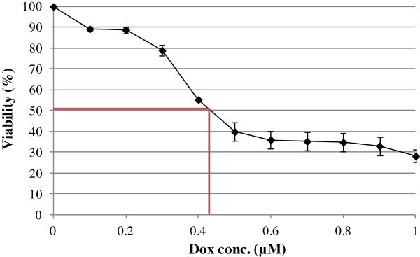
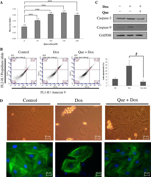
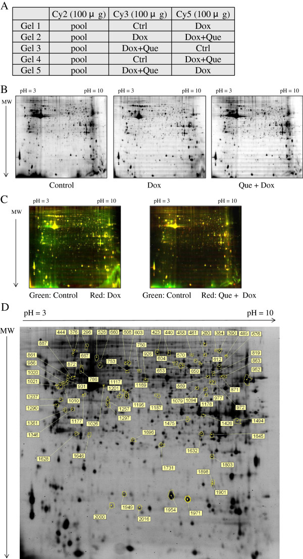
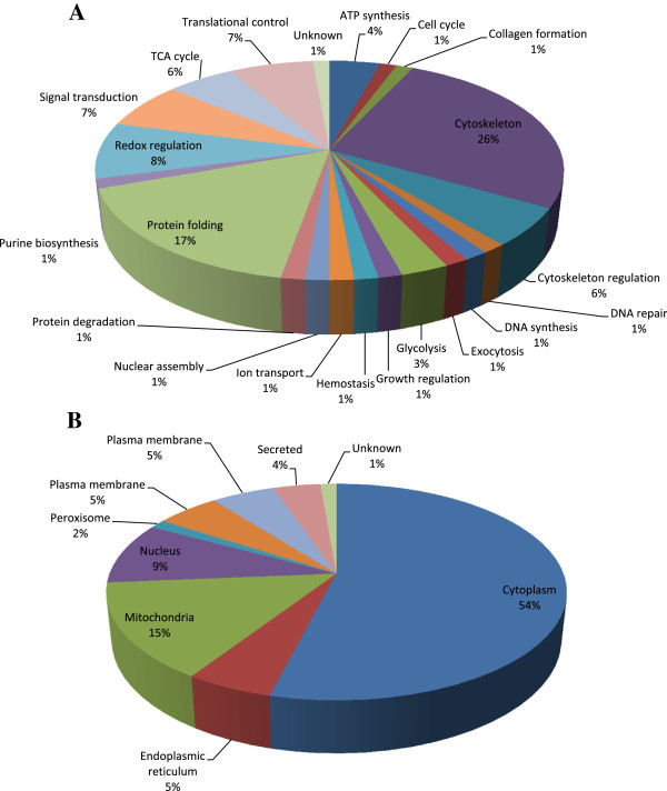
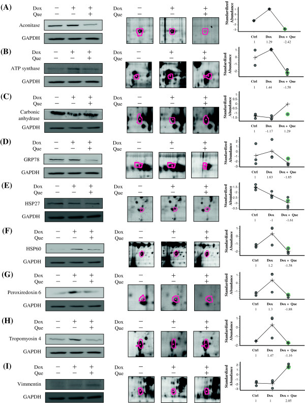
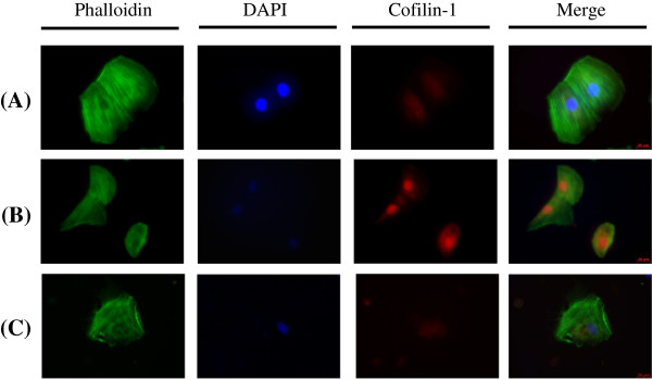
Results: Proteomic analysis and a cell biology assay were performed to investigate the quercetin-induced responses. Our data demonstrated that quercetin treatment protects the cardiomyocytes in a doxorubicin-induced heart damage model. Quercetin significantly facilitated cell survival by inhibiting cell apoptosis and maintaining cell morphology by rearranging the cytoskeleton. Additionally, 2D-DIGE combined with MALDI-TOF MS analysis indicated that quercetin might stimulate cardiomyocytes to repair damage after treating doxorubicin by modulating metabolic activation, protein folding and cytoskeleton rearrangement.

Conclusion: Based on a review of the literature, this study is the first to report detailed protective mechanisms for the action of quercetin against doxorubicin-induced cardiomyocyte toxicity based on in-depth cell biology and proteomic analysis.


Figures

Figure 1
Figure 1
Effect of doxorubicin treatment on H9C2 cell viability. H9C2 cells were treated with indicated concentrations of doxorubicin from 3 independent experiments. Cell viability was determined by MTT assay after 24 h exposure of doxorubicin. Each data point indicates mean ± SD of triplicate values.
Figure 2
Figure 2
Effects of quercetin on doxorubicin-induced changes of cell viability, cell apoptosis and cell morphology in H9C2 cells. (A) MTT-based viability assays were performed on H9C2 cell cultures following treatments with different concentrations of quercetin (50 μM, 100 μM, 150 μM and 200 μM) or left untreated. Values were normalized against untreated samples and were the average of 4 independent measurements +/- the standard deviation. The statistic analysis was performed with two group paired Student t-test. (B) Typical dot plot diagrams detected annexin V-FITC and PI staining represent untreated, doxorubicin-treated, and quercetin-pretreated followed by doxorubicin–treated cells. The x-axis and y-axis stand for the intensity of annexin V-FITC and PI, respectively. The lower left area of presented background staining by annexin V-FITC and PI in normal cells, and apoptotic signals located in the right area. This figure is representative of 4 replicates. The statistic analysis of the replicates was listed in right panel. (C) The levels of caspase 3 and caspase 9 in H9C2 cells were detected by immunoblotting. GAPDH served as a sample loading control. (D) Cell morphology and protein location of F-actin in H9C2 cells were analyzed by immunostaining. H9C2 cells on coverslips were either left untreated, treated with doxorubicin or pre-treated with quercetin prior to doxorubicin treatment before fixation and staining. F-actin was stained with phalloidin and nuclei were stained with DAPI. Each set of five fields were taken using the same exposure and images are representative of five different fields. In (B) ~ (D), H9C2 cells were untreated, 0.45 μM of doxorubicin for 24 h, or 100 μM of quercetin for 4 h followed by 0.45 μM of doxorubicin for 24 h.
Figure 3
Figure 3
2D-DIGE analysis of H9C2 cells in response to doxorubicin treatment and pre-treatment with quercetin. (A) Samples arrangement for a triplicate 2D-DIGE experiment. (B) Protein samples (100 μg each) were labeled with Cy-dyes and separated using 24 cm, pH 3-10 non-linear IPG strips. 2D-DIGE images of the protein samples from H9C2 cells in response to doxorubicin treatment and pre-treatment with quercetin at appropriate excitation and emission wavelengths were shown as well as overlaid pseudo-colored images processed with ImageQuant Tool (GE Healthcare) (C). (D) Protein samples (100 μg each) purified from total cell lysates were labeled with Cy-dyes and separated using 24 cm, pH 3-10 non-linear IPG strips. The differentially expressed protein features were annotated with spot numbers. In this 2D-DIGE experiment, H9C2 cells were untreated, 0.45 μM of doxorubicin for 24 h, or 100 μM of quercetin for 4 h followed by 0.45 μM of doxorubicin for 24 h.
Figure 4
Figure 4
Percentage of total differentially expressed proteins identified by 2D-DIGE/MALDI-TOF MS for H9C2 cells in response to doxorubicin treatment and pre-treatment with quercetin according to their biological functions (A) and sub-cellular locations (B).
Figure 5
Figure 5
Representative immunoblotting and immunofluorescent analyses for selected differentially expressed proteins identified by proteomic analysis of H9C2 cells in response to doxorubicin treatment and pre-treatment with quercetin. The levels of identified proteins, (A) Aconitase, (B) ATP synthase, (C) Carbonic anhydrase, (D) GRP78, (E) HSP27, (F) HSP60, (G) Peroxiredoxin 6, (H) Tropomyosin 4, (I) Vimmentin, in H9C2 cells in response to doxorubicin treatment and pre-treatment with quercetin were confirmed by immunoblot, while GAPDH was used as loading controls (left panels). The protein expression maps and two-dimensional spot images were shown in right panels and middle panels, respectively. (J) H9C2 cells in different treatment conditions were fixed and incubated with anti-Cofilin-1 antibody (Red) and stained with Phalloidin (Green). Nucleus were stained with DAPI (Blue). Each set of three fields was taken using the same exposure, and images are representative of three different fields. In this validation experiment, H9C2 cells were left untreated, treated with 0.45 μM doxorubicin for 24 h or pretreated with 100 μM quercetin for 4 h followed by 0.45 μM doxorubicin for further 24 h.
Figure 6
Figure 6
Representative immunofluorescent analyses for Cofilin identified by proteomic analysis of H9C2 cells in response to doxorubicin treatment and pre-treatment with quercetin.H9C2 cells in different treatment conditions were fixed and incubated with anti-Cofilin-1 antibody (Red) and stained with Phalloidin (Green). Nucleus was stained with DAPI (Blue). Each set of three fields was taken using the same exposure, and images are representative of three different fields. In this validation experiment, H9C2 cells were left untreated, treated with 0.45 µM doxorubicin for 24 h or pretreated with 100 µM quercetin for 4 h followed by 0.45 µM doxorubicin for further 24 h.

References

    1. Verma S, Dent S, Chow BJ, Rayson D, Safra T. Metastatic breast cancer: the role of pegylated liposomal doxorubicin after conventional anthracyclines. Cancer Treat Rev. 2008;20:391–406. doi: 10.1016/j.ctrv.2008.01.008. - DOI - PubMed
    1. Vatsyayan R, Chaudhary P, Lelsani PC, Singhal P, Awasthi YC, Awasthi S, Singhal SS. Role of RLIP76 in doxorubicin resistance in lung cancer (Review) Int J Oncol. 2009;20:1505–1511. - PMC - PubMed
    1. Green AE, Rose PG. Pegylated liposomal doxorubicin in ovarian cancer. Int J Nanomedicine. 2006;20:229–239. - PMC - PubMed
    1. Christiansen S, Autschbach R. Doxorubicin in experimental and clinical heart failure. Eur J Cardiothorac Surg. 2006;20:611–616. doi: 10.1016/j.ejcts.2006.06.024. - DOI - PubMed
    1. Kalishina EV, Saprin AN, Solomka VS, Shchebrak NP, Piruzian LA. Inhibition of hydrogen peroxide, oxygen and semiquinone radicals in the development of drug resistance to doxorubicin in human erythroleukemia K562-cells. Vopr Onkol. 2003;20:294–298. - PubMed