. 2014 May;34(5):394-403.
doi: 10.1089/jir.2013.0062. Epub 2013 Dec 20.

High-dose interferon-γ promotes abortion in mice by suppressing Treg and Th17 polarization

Affiliations

High-dose interferon-γ promotes abortion in mice by suppressing Treg and Th17 polarization

Hai-Yan Liu et al. J Interferon Cytokine Res. 2014 May.

Abstract

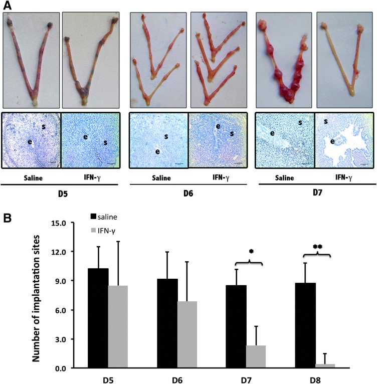
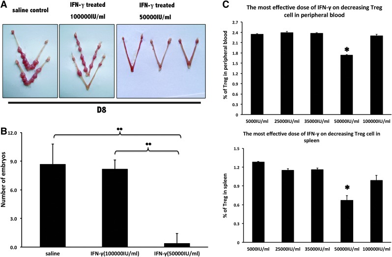
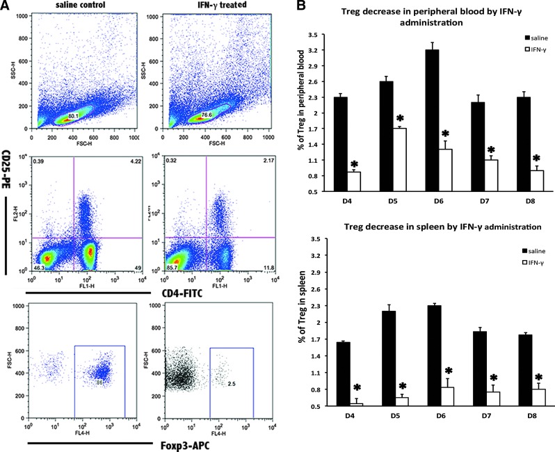
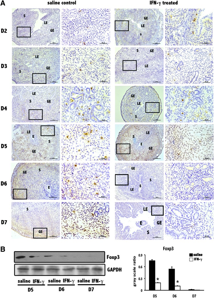
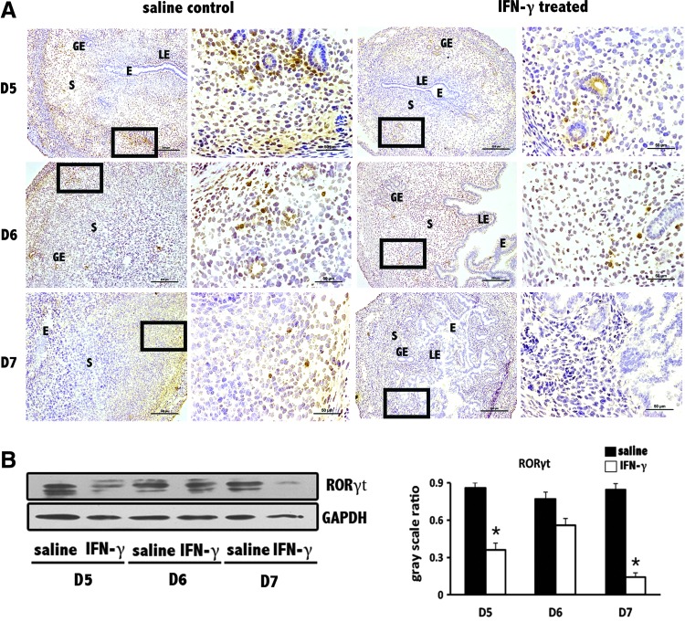
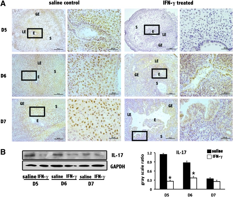
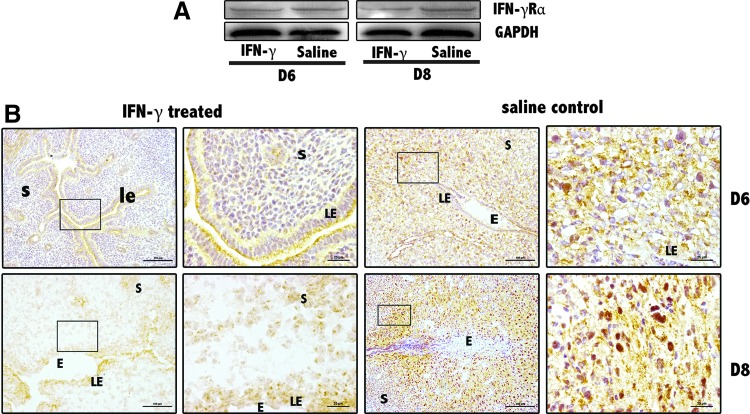
As a classic type I cytokine, interferon-gamma (IFN-γ) is known to manifest a miscarriage-inducing effect, although the specific mechanism is still unclear. To determine whether immune cells such as regulatory T (Treg) and Th17 cells are involved in these abortions, syngeneically pregnant (BALB/c×BALB/c) mice were subjected to intravaginal IFN-γ administration (5 × 10(3) IU/mouse on D3 of gestation). These mice experienced significant fetal loss on D7/D8 of pregnancy, and a remarkable drop in the Treg cell ratio was observed in the peripheral blood and the spleen by flow cytometry. In situ detection of the uterine tissue peri-implantation revealed that IFN-γ treatment also caused statistically significant reductions in forkhead box P3, RAR-related orphan receptor gamma, and IL-17 levels, which indicated local decreases in Treg and Th17 cells at uterine implantation sites. The IFN-γ receptor alpha (IFN-γRα) level was also lowered in the uterus. These results demonstrate that in murine pregnancy, a supraphysiological dose of IFN-γ could induce peri-implantation failure. Moreover, in this study, the decreases in both Treg and Th17-type cells, which may be relevant to the role of IFN-γRα, may be one of the main reasons that IFN-γ causes abortion.


Figures

<b>FIG. 1.</b>
FIG. 1.
The miscarriage-inducing effect of interferon-gamma (IFN-γ) in mice peri-implantation (D4–D7 of pregnancy). (A) Overall pregnancy uterine pictures and representative uterine implantation site sections. Trypan blue was administered by mouse tail vein injection to stain the implantation sites of the uterus on D5. The uterine implantation site sections were stained with hematoxylin. The letter e is for embryo and s is for uterine stroma. (B) Histogram of implantation site numbers in the uterus on D5–D7 with the administration of IFN-γ. All results are expressed in terms of the implantation site number in the uterus on different days of pregnancy. The bars represent the SD of the mean. The data at each point were derived from 9 separate samples from pregnant mice. A total of 54 samples from pregnant mice were observed to perform this count. *P<0.05, **P<0.01.
<b>FIG. 2.</b>
FIG. 2.
Determination of the most effective miscarriage-inducing dose of mIFN-γ. (A) Overall pregnancy uterine pictures on D8 of pregnancy. (B) Histogram of implantation site numbers in the uterus on D8 with the administration of IFN-γ at different doses. The data at each point were derived from at least 7 separate samples from pregnant mice. **P<0.01. (C) Effects of different doses of IFN-γ on the regulatory T (Treg) cell ratios in the peripheral blood and the spleen. All results are expressed in terms of the % of Treg cells (CD4+CD25+Foxp3+) at different IFN-γ doses. The bars represent the SEM. One-way ANOVA was used to assess the significance of differences. *P<0.05. *Also showed significant differences between the dose of 50,000 IU/mL and other doses (5,000, 25,000, 35,000, and 100,000 IU/mL). There was no significant difference between other compared groups (P>0.05).
<b>FIG. 3.</b>
FIG. 3.
Treg cell decrease in the peripheral blood and the spleen following IFN-γ injection into mice peri-implantation. (A) Flow cytometry scatterplot of Treg cells. Treg cells are CD4+CD25+Foxp3+. (B) The Treg cell proportion decreased significantly in the peripheral blood and the spleen under the influence of supraphysiological dose of mIFN-γ. The CD4+CD25+Foxp3+ Treg cell ratio was evaluated by flow cytometry analysis. The data are expressed as the percent of CD4+CD25+Foxp3+ triple-positive cells. All mIFN-γ administrations were performed at a dose of 5×10⁴ IU/mL, with 100 μL per mouse. The bars represent the SD of the mean. A paired t-test was used to assess the significance of differences. *P<0.05, IFN-γ treatment group versus the corresponding saline control group.
<b>FIG. 4.</b>
FIG. 4.
Decrease in forkhead box P3 (Foxp3) levels at uterine implantation sites. (A) Foxp3 levels were obviously reduced at implantation sites based on immunohistochemistry. This signal reduction was not obvious on D2 but became more and more remarkable as the pregnancy progresses. Scale bars=200 and 50 μm. At least 3 independent experiments were repeated for each time point. The data at each point were derived from 3 separate samples from pregnant mice. A total of 40 samples from pregnant mice were assessed. (B) Foxp3 levels were significantly reduced at uterine implantation sites based on Western blotting. GAPDH was used as a loading control. The bars represent the SD of the mean of the relative value in gray (Foxp3/GAPDH). A paired t-test was used to assess the significance of differences. *P<0.05, IFN-γ treatment group versus the corresponding saline control group. At least 3 independent experiments were repeated for each time point.
<b>FIG. 5.</b>
FIG. 5.
Decrease in RAR-related orphan receptor gamma (RORγt) levels at uterine implantation sites. (A) RORγt levels were obviously reduced at implantation sites based on immunohistochemistry. Scale bars=200 and 25 μm. At least 3 independent experiments were repeated for each time point. The data at each point were derived from 3 separate samples from pregnant mice. A total of 18 samples from pregnant mice were assessed. (B) RORγt levels were significantly reduced at uterine implantation sites based on Western blotting. GAPDH was used as a loading control. The bars represent the SD of the mean of the relative value in gray (RORγt/GAPDH). A paired t-test was used to assess the significance of differences. *P<0.05, IFN-γ treatment group versus the corresponding saline control group. At least 3 independent experiments were repeated for each time point.
<b>FIG. 6.</b>
FIG. 6.
Decrease in IL-17 levels at uterine implantation sites. (A) IL-17 levels were obviously reduced at implantation sites based on immunohistochemistry. Scale bars=200 and 50 μm. At least 3 independent experiments were repeated for each time point. The data at each point were derived from 3 separate samples from pregnant mice. A total of 18 samples from pregnant mice were assessed. (B) IL-17 levels were significantly reduced at implantation sites based on Western blotting. GAPDH was used as a loading control. The bars represent the SD of the mean of the relative value in gray (IL-17/GAPDH). A paired t-test was used to assess the significance of differences. *P<0.05, IFN-γ treatment group versus the corresponding saline control group. At least 3 independent experiments were repeated for each time point.
<b>FIG. 7.</b>
FIG. 7.
Decrease in IFN-γ receptor alpha (IFN-γRα) levels at uterus implantation sites. (A) IFN-γRα levels were significantly reduced at D6 and D8 implantation sites based on Western blotting. GAPDH was used as a loading control. At least 3 independent experiments were repeated for each time point. (B) IFN-γRα levels were obviously reduced at implantation sites based on immunohistochemistry. Scale bars=100 and 25 μm. At least 3 independent experiments were repeated for each time point. The data at each point were derived from 3 separate samples from pregnant mice. A total 12 samples from pregnant mice were assessed.

References

    1. Aluvihare VR, Kallikourdis M, Betz AG. 2004. Regulatory T cells mediate maternal tolerance to the fetus. Nat Immunol 5(3):266–271 - PubMed
    1. Arruvito L SM, Banham AH, Fainboim L. 2007. Expansion of CD4+CD25+ and FOXP3+ regulatory T cells during the follicular phase of the menstrual cycle: implications for human reproduction. J Immunol 178(4):2572–2578 - PubMed
    1. Basta P, Majka M, Jozwicki W, Lukaszewska E, Knafel A, Grabiec M, Stasienko E, Wicherek L. 2010. The frequency of CD25+CD4+ and FOXP3+ regulatory T cells in ectopic endometrium and ectopic decidua. Reprod Biol Endocrinol 8:116. - PMC - PubMed
    1. Chaouat G, Assal Meliani A, Martal J, Raghupathy R, Elliott JF, Mosmann T, Wegmann TG. 1995. IL-10 prevents naturally occurring fetal loss in the CBA X DBA/2 mating combination, and local defect in IL-10 production in this abortion-prone combination is corrected by in vivo injection of IFN-tau. J Immunol 154(9):4261–4268 - PubMed
    1. Chaouat G, Menu E, Clark DA, Dy M, Minkowski M, Wegmann TG. 1990. Control of fetal survival in CBA X DBA/2 mice by lymphokine therapy. J Reprod Fertil 89(2):447–458 - PubMed

Publication types

MeSH terms