Review
. 2013 Dec 17;105(12):2621-8.
doi: 10.1016/j.bpj.2013.10.032.

Regulation of actin by ion-linked equilibria

Affiliations
Review

Regulation of actin by ion-linked equilibria

Hyeran Kang et al. Biophys J. .

Abstract

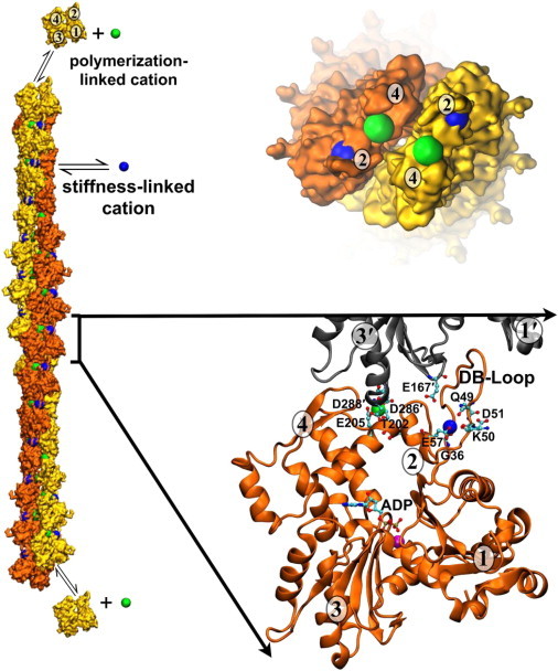
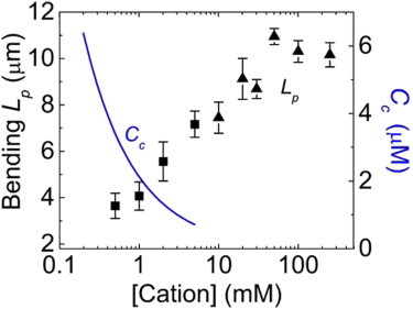
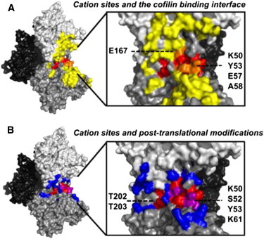
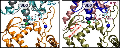
Actin assembly, filament mechanical properties, and interactions with regulatory proteins depend on the types and concentrations of salts in solution. Salts modulate actin through both nonspecific electrostatic effects and specific binding to discrete sites. Multiple cation-binding site classes spanning a broad range of affinities (nanomolar to millimolar) have been identified on actin monomers and filaments. This review focuses on discrete, low-affinity cation-binding interactions that drive polymerization, regulate filament-bending mechanics, and modulate interactions with regulatory proteins. Cation binding may be perturbed by actin post-translational modifications and linked equilibria. Partial cation occupancy under physiological and commonly used in vitro solution conditions likely contribute to filament mechanical heterogeneity and structural polymorphism. Site-specific cation-binding residues are conserved in Arp2 and Arp3, and may play a role in Arp2/3 complex activation and actin-filament branching activity. Actin-salt interactions demonstrate the relevance of ion-linked equilibria in the operation and regulation of complex biological systems.


Figures

Figure 1
Figure 1
Location of two discrete, actin filament-specific cation-binding sites. The actin filament structure is based on the model of Fujii et al. (50) and includes the predicted cation-binding sites from Kang et al. (18). The barbed end of the filament is toward the bottom of the figure and the pointed end with associated cations is shown following a 90° rotation. (Yellow and orange) Surface rendering of actin monomers; (orange and gray) cartoon rendering of actin monomers. (Numbers in ovals) Actin monomer subdomains. (Green) Polymerization site cation; (blue) stiffness site cation; (magenta) nucleotide-associated cation. (Ball-and-stick representation) ADP nucleotide and specific cation-binding residues. To see this figure in color, go online.
Figure 2
Figure 2
Two distinct classes of cation interaction sites exist on actin. One class responds to submillimolar cation concentrations and lowers Cc (blue; data shown for Mg2+). The second class responds to millimolar concentration and stiffens filaments as measured by Lp (black; squares represent Mg2+, triangles represent K+). Figure adapted from Kang et al. (18). To see this figure in color, go online.
Figure 3
Figure 3
Actin regulatory protein-binding interfaces and posttranslational modification sites overlap with filament-specific cation sites. (A) Residues participating in the actin-cofilin interface (yellow) (78); (inset) 90° turn and zoom to cation sites where site residues in the cofilin binding interface are highlighted (orange). (B) Acetylation, ADP-ribosylation, arginylation, carbonylation, malonylation, methylation, nitrosylation, oxidation, phosphorylation, and ubiquitylation sites (83) are shown (blue) for longitudinal subunits; (inset) 90° turn and zoom to cation sites where modifiable site residues are highlighted (purple). To see this figure in color, go online.
Figure 4
Figure 4
Conserved cation-binding residues between Arp2/3 and actin suggest salt-dependent regulation of Arp2/3 activation. Arp2 and Arp3 are overlaid with an actin subunit (best alignment calculated by the software FATCAT (88)) and interact with the next longitudinally neighboring subunit at the barbed-end face of Arp2/3. Actin subunits at the bottom of each panel represent the first two actin monomers that associate with Arp2/3 to nucleate the daughter filament at a branch point. Arp2 aligns very well with actin, especially the putative cation-binding residues E171, D290, and D292, which help form the polymerization and stiffness cation-binding sites with the incoming actin monomer. Arp3 does not align as well in the inactive crystal conformation. Arp2/3 activation is thought to require WASP/Scar-dependent conformational rearrangement of Arp3. We hypothesize that this rearrangement relieves a steric clash with the incoming actin monomer while forming a better cation-binding geometry at both the polymerization and stiffness cation-binding sites shared with the incoming daughter filament subunit. In this figure, pivoting of the Arp3 SD3 to the left would both alleviate the steric clash and place E182 and D310 into the proper position to bind interfacial cations. To see this figure in color, go online.

References

    1. Korn E.D., Carlier M.F., Pantaloni D. Actin polymerization and ATP hydrolysis. Science. 1987;238:638–644. - PubMed
    1. Pollard T.D., Borisy G.G. Cellular motility driven by assembly and disassembly of actin filaments. Cell. 2003;112:453–465. - PubMed
    1. Pollard T.D., Cooper J.A. Actin, a central player in cell shape and movement. Science. 2009;326:1208–1212. - PMC - PubMed
    1. Pollard T.D., Blanchoin L., Mullins R.D. Molecular mechanisms controlling actin filament dynamics in nonmuscle cells. Annu. Rev. Biophys. Biomol. Struct. 2000;29:545–576. - PubMed
    1. Ditlev J.A., Mayer B.J., Loew L.M. There is more than one way to model an elephant. Experiment-driven modeling of the actin cytoskeleton. Biophys. J. 2013;104:520–532. - PMC - PubMed

Publication types

LinkOut - more resources