. 2013 Dec 20:6:58.
doi: 10.1186/1756-6606-6-58.

α7nAchR/NMDAR coupling affects NMDAR function and object recognition

Affiliations

α7nAchR/NMDAR coupling affects NMDAR function and object recognition

Shupeng Li et al. Mol Brain. .

Abstract

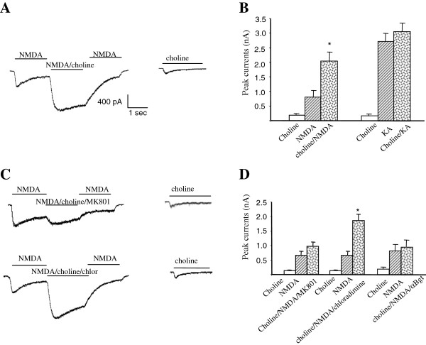
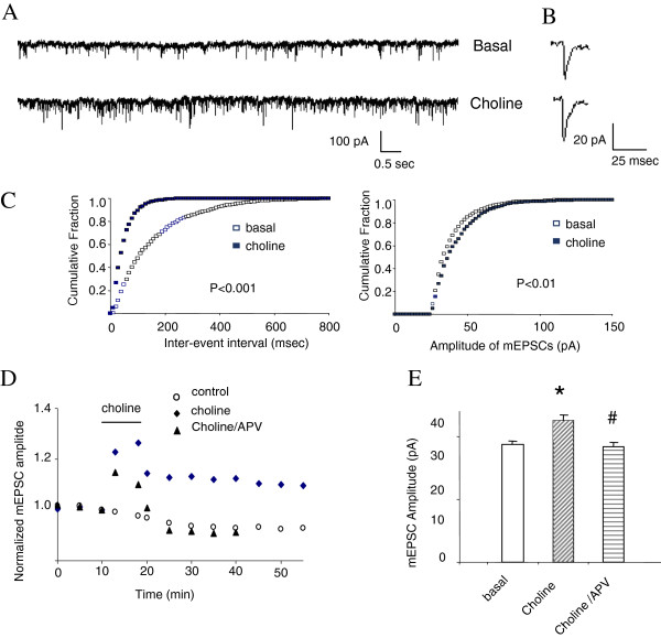
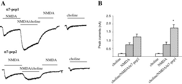
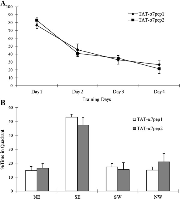
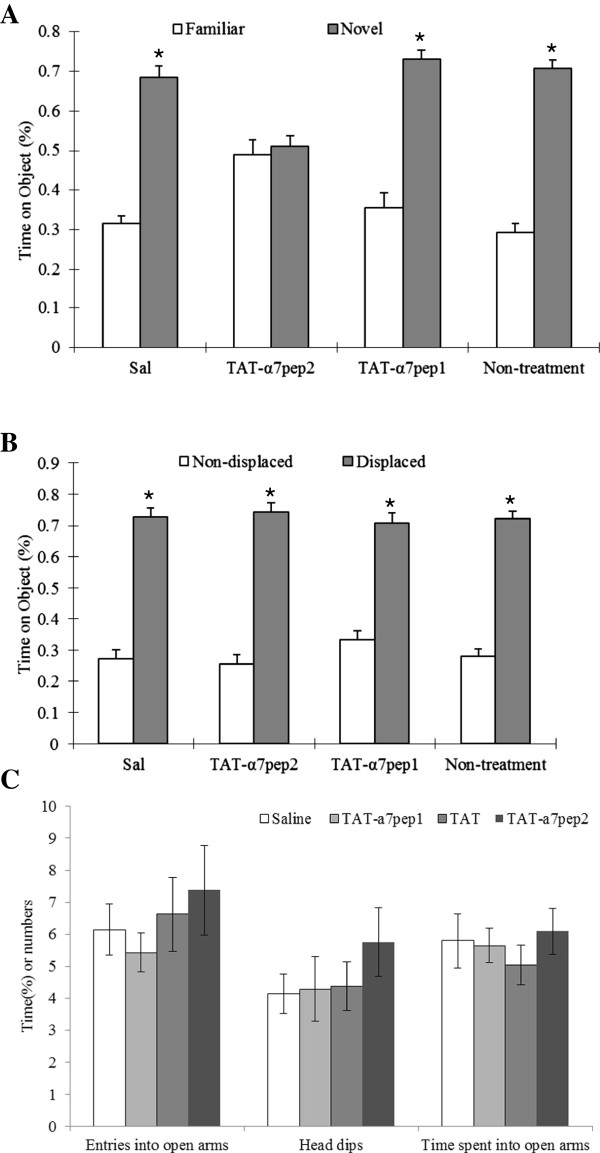
The α7 nicotinic acetylcholine receptor (nAchR) and NMDA glutamate receptor (NMDAR) are both ligand-gated ion channels permeable to Ca2+ and Na+. Previous studies have demonstrated functional modulation of NMDARs by nAchRs, although the molecular mechanism remains largely unknown. We have previously reported that α7nAchR forms a protein complex with the NMDAR through a protein-protein interaction. We also developed an interfering peptide that is able to disrupt the α7nAchR-NMDAR complex and blocks cue-induced reinstatement of nicotine-seeking in rat models of relapse. In the present study, we investigated whether the α7nAchR-NMDAR interaction is responsible for the functional modulation of NMDAR by α7nAchR using both electrophysiological and behavioral tests. We have found that activation of α7nAchR upregulates NMDAR-mediated whole cell currents and LTP of mEPSC in cultured hippocampal neurons, which can be abolished by the interfering peptide that disrupts the α7nAchR-NMDAR interaction. Moreover, administration of the interfering peptide in mice impairs novel object recognition but not Morris water maze performance. Our results suggest that α7nAchR/NMDAR coupling may selectively affect some aspects of learning and memory.


Figures

Figure 1
Figure 1
Choline induced synergistic effect on NMDAR currents through the α 7-nAchR/NMDAR direct protein-protein interaction. (A) Co-application of 1 mM choline with 50 µM NMDA/10 µM glycine produced a synergistic effect that display a significantly larger current compared to the current induced by NMDA/Glycine alone (n = 43 of 47 cells, P < 0.01). (B) The choline induced synergistic effect is specific to NMDAR-mediated currents since no such an effect was detected on currents induced by 100 µM kainic acid. (C, D) The choline-induced synergistic effect is significantly inhibited by simultaneous application NMDAR channel blocker MK-801 (10 µM) (n = 8, p < 0.05), but not the nAchR channel blocker chlorisondamine (20 µM). Furthermore, pretreatment of the neurons with the α7nAchR specific antagonist α-Bungarotoxin for 40 minutes inhibited the choline-induced synergistic effect.
Figure 2
Figure 2
Choline induced upregulation of NMDAR-dependent LTP of mEPSCs in cultured hippocampal neurons. (A) Examples of continuous recordings from individual neurons 5 minutes before (Basal) and 30 minutes after 8-minute stimulation of neurons with 1 mM choline. (B) Single events taken from the basal and choline traces, respectively showing that the amplitude of mEPSCs was increased by choline application. (C) Cumulative fraction plots for mEPSCs inter-event intervals and amplitudes obtained 5 minutes before (Basal) and 30 minutes after choline (8 min, 1 mM). (D) mEPSC amplitudes are normalized to the values from the initial 10 min and plotted over time. Treatment of neurons with choline (8 min, 1 mM) significantly increased the amplitude of the mEPSCs over the time course of recordings; an effect can be abolished by NMDAR antagonist, AP5 (100 μM). (E) Amplitude histogram summarizes data from groups of individual neurons treated with glycine (200 μM; 3 min) in the absence or presence of choline (1 mM) or choline/AP5 (100 μM). Responses obtained 30 min after glycine treatment (26.5+/- 2.3 pA), 30 min after choline treatment (31.4 +/-2.7 pA, n = 6, *p < 0.01) and 30 minutes after coapplication of choline/APV (25.9+/-2.0pA n = 3, **p < 0.05, paired t-test).
Figure 3
Figure 3
Application of α7pep2 peptide blocked choline induced upregulation of NMDA current in hippocampal primary culture. (A) The choline-induced synergistic effect is significantly inhibited by intracellular application of interfering peptides α7pep2, but not α7pep1 (10 µM) (choline/NMDA: 1202.7 ± 182.1 pA; NMDA: 910.5 ± 130.8, n = 6, p > 0.05). Cells were hold at -70 mV, 20 mM bicuculline, 1 mM strychnine, 0.5 μM TTX, 1 mM glycine were included in the extracellular solution. (B) Amplitude histogram summarizes data from groups of individual neurons treated with glycine (200 μM; 3 min) in the absence or presence of choline (1 mM) with the intracellular application of α7pep1, α7pep2 peptide respectively. Responses obtained 30 min after glycine treatment (basal) and 30 min after choline treatment (choline). α7pep1 peptide did not block the enhancing effect of choline on the mEPSC amplitude (basal: 25.2 +/-2.1 pA; choline: 28.4+/-2.4 pA, n = 4, *p < 0.01, paired t-test) while choline failed to upregulate mEPSC amplitude with the presence of α7pep2 (basal: 24.2+/-2.0; choline: 25.1 +/-2.3 pA, n = 6, p > 0.05, paired t-test).
Figure 4
Figure 4
Application of α7pep2 peptide blocked choline induced upregulation of mEPSC of LTP in hippocampal primary culture. (A) Examples of continuous recordings from individual neurons 40 minute after intracellular application of α7pep2 peptide (10 µM) with/without the presence of choline (1 mM, 8 min). (B) Single events taken from the basal and choline traces after intracellular application of α7pep2 peptide, showing that choline application failed to increase the amplitude of mEPSCs. (C) Cumulative fraction plots for mEPSCs inter-event intervals and amplitudes obtained 5 minutes before (Basal) and 30 minutes after choline (8 min, 1 mM) with the presence of α7pep2 peptide intracellularly. (D) Amplitude histogram summarizes data from groups of individual neurons treated with glycine (200 μM; 3 min) in the absence or presence of choline (1 mM) with the intracellular application of α7pep1, α7pep2 peptide respectively. Responses obtained 30 min after glycine treatment (basal) and 30 min after choline treatment (choline). α7pep1 peptide did not block the enhancing effect of choline on the mEPSC amplitude (basal: 25.2 +/-2.1 pA; choline: 28.4+/-2.4 pA, n = 4, *p < 0.01, paired t-test) while choline failed to upregulate mEPSC amplitude with the presence of α 7pep2 (basal: 24.2+/-2.0; choline: 25.1 +/-2.3 pA, n = 6, p > 0.05, paired t-test).
Figure 5
Figure 5
TAT-α7pep2 peptide treatment has no effects on spatial learning and memory. Latency to find the platform of mice was not affected by peptide treatment in the Morris water maze task. (A) In the acquisition phase, escape latency to find a hidden platform located in the southeast (SE) quadrant was unaffected by treatment. (B) Histogram of percent time spent in each quadrant at probe test.
Figure 6
Figure 6
TAT-α7pep2 Peptide treatment affected nonspatial learning and memory. TAT-α7pep2 Peptide treatment impaies novel object recognition (A) but not displaced object recognition (B). Times of exploration of the DO and NDO were recorded and expressed as a percentage of the total time of objects investigated. In the novel object recognition session, one of the familiar NDOs was replaced with a new object (NO) at the same location and the two familiar DOs were removed. Data were analyzed with ANOVA with treatment as a between-subjects factor, and object rearrangement or object replacement as a repeated measures factor. The Tukey test was used for post hoc comparisons when ANOVA yielded statistically significant main effects or interactions. (C) In the elevated plus maze, no significant changes in the percent of time spent in open arms, entries into the open arms and head dips were observed in different treatment groups.

References

    1. Bartlett TE, Wang YT. The intersections of NMDAR-dependent synaptic plasticity and cell survival. Neuropharmacology. 2013;6:59–68. - PubMed
    1. Bliss TV, Collingridge GL. Expression of NMDA receptor-dependent LTP in the hippocampus: bridging the divide. Mol Brain. 2013;6:5. doi: 10.1186/1756-6606-6-5. - DOI - PMC - PubMed
    1. Paoletti P, Bellone C, Zhou Q. NMDA receptor subunit diversity: impact on receptor properties, synaptic plasticity and disease. Nat Rev Neurosci. 2013;6(6):383–400. doi: 10.1038/nrn3504. - DOI - PubMed
    1. Gray R, Rajan AS, Radcliffe KA, Yakehiro M, Dani JA. Hippocampal synaptic transmission enhanced by low concentrations of nicotine. Nature. 1996;6(6602):713–716. doi: 10.1038/383713a0. - DOI - PubMed
    1. MacDermott AB, Mayer ML, Westbrook GL, Smith SJ, Barker JL. NMDA-receptor activation increases cytoplasmic calcium concentration in cultured spinal cord neurones. Nature. 1986;6(6069):519–522. doi: 10.1038/321519a0. - DOI - PubMed

Publication types

MeSH terms

LinkOut - more resources