. 2013 Dec 19;155(7):1581-1595.
doi: 10.1016/j.cell.2013.10.056.

Brd4 and JMJD6-associated anti-pause enhancers in regulation of transcriptional pause release

Affiliations

Brd4 and JMJD6-associated anti-pause enhancers in regulation of transcriptional pause release

Wen Liu et al. Cell. .

Abstract

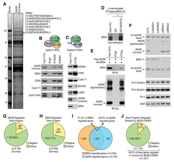
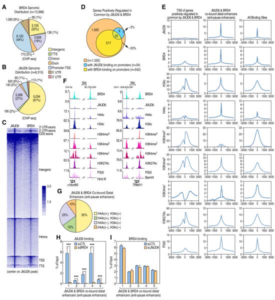
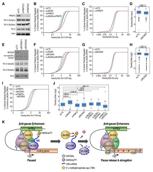
Distal enhancers characterized by the H3K4me(1) mark play critical roles in developmental and transcriptional programs. However, potential roles of specific distal regulatory elements in regulating RNA polymerase II (Pol II) promoter-proximal pause release remain poorly investigated. Here, we report that a unique cohort of jumonji C-domain-containing protein 6 (JMJD6) and bromodomain-containing protein 4 (Brd4) cobound distal enhancers, termed anti-pause enhancers (A-PEs), regulate promoter-proximal pause release of a large subset of transcription units via long-range interactions. Brd4-dependent JMJD6 recruitment on A-PEs mediates erasure of H4R3me(2(s)), which is directly read by 7SK snRNA, and decapping/demethylation of 7SK snRNA, ensuring the dismissal of the 7SK snRNA/HEXIM inhibitory complex. The interactions of both JMJD6 and Brd4 with the P-TEFb complex permit its activation and pause release of regulated coding genes. The functions of JMJD6/ Brd4-associated dual histone and RNA demethylase activity on anti-pause enhancers have intriguing implications for these proteins in development, homeostasis, and disease.


Figures

Figure 1
Figure 1. Physical Interaction between JMJD6 and Brd4 in the Context of Active P-TEFb Complex
(A) JMJD6 associated proteins were purified using Flag-affinity agarose, separated by SDS-PAGE gel, stained with Coomassie blue, and then subjected to mass spectrometry analysis. Identified peptides for Brd4 were shown on the right. (B) Nuclear extracts purified from HEK293T cells were subjected to immunoprecipitation (IP) and followed by immunoblotting (IB). (C) In vitro 7SK RNA pull-down assay was performed and the resultant pellet was analyzed by IB. (D, E) JMJD6 and Brd4 interaction examined through in vitro GST pull-down assay (D) or IP in HEK293T cells (E). Note that JMJD6 undergoes multimerization, which is preserved even under denatured conditions. (F) HEK293T cells transfected with control siRNA or two independent siRNAs specific targeting JMJD6 or Brd4 were subjected to IB analysis. (G, H) Pie chart showing genes regulated by JMJD6 (G) or Brd4 (H) assessed by Gro-seq analysis in cells as described in (F) (FDR<0.001). (I) Overlap between JMJD6 and Brd4 regulated genes from (G) and (H), respectively. (J) Genes regulated in common and in the same direction by JMJD6 and Brd4.
Figure 2
Figure 2. JMJD6 and Brd4 Promote Promoter-proximal Pol II Pause Release
(A) Schematic representation of the way applied to calculate Pol II traveling ratio (TR). (B, D) Pol II TR distribution in HEK293T cells for all Pol II-bound genes (B) or genes that are positively-regulated in common by JMJD6 and Brd4 (n=1,022) (JB genes) (D). (C, E) Box plots showing the change of Pol II TR caused by knock-down of JMJD6 or Brd4 in panel B (C) and D (E) (Median (C): 6.54 (siCTL), 10.36 (siBrd4), 7.48 (siJMJD6); Median (E): 2.61 (siCTL), 5.46 (siBrd4), 4.14 (siJMJD6)). (F) Gro-seq read distribution along the transcription units from 2kb upstream of the transcription start site (TSS) to 2.5kb downstream of the transcription termination site (TTS) in HEK293T cells transfected with control, JMJD6 or Brd4 siRNA. Region included was normalized and scaled to 1. (G, I) TR distribution based on Gro-seq analysis in HEK293T cells for all Pol II-bound genes (G) or JB genes (I). (H, J) Box plots showing change of TR caused by knock-down of JMJD6 or Brd4 as shown in panel G (H) and I (J) (Median (H): 0.61 (siCTL), 1.82 (siBrd4), 1.00 (siJMJD6); Median (J): 0.33 (siCTL), 1.22 (siBrd4), 0.66 (siJMJD6)).
Figure 3
Figure 3. JMJD6 and Brd4 Genomic Binding
(A, B) Genomic distribution of Brd4 (A) or JMJD6 (B) binding sites. (C) Heat map representation of JMJD6 and Brd4 tag density centered on JMJD6 ChIP-seq peaks. (D) Venn diagram showing the number of genes having JMJD6 and/or Brd4 binding on promoter regions among those JB genes. (E) ChIP-seq tag distribution of JMJD6, Brd4, H3Ac, H4Ac, H3K4me3, H3K4me2, H3K4me1, H3K27Ac or P300 surrounding TSS of those JB genes (left panel), the center of JMJD6 and Brd4 co-bound distal sites (middle panel) or all of its own binding sites in the genome (right panel). (F) JMJD6, Brd4, H3Ac, H4Ac, H3K4me3, H3K4me2, H3K4me1, H3K27Ac or P300 binding on selected genomic regions: C16orf93 or TRIM11 promoter region and the nearby JMJD6 and Brd4 co-bound distal enhancer (Center of enhancer: chr16:30,733,200 (left panel); chr1:226,633,061 (right panel)). (G) Pie chart displaying the percentage of JMJD6 and Brd4 co-bound distal sites with or without H4Ac or H3Ac marks. (H, I) Brd4 is required for JMJD6 binding on chromatin, but not vice versa. Standard ChIP assay was performed with anti-JMJD6 (H) or anti-Brd4 (I) antibody in HEK293T cells. The five regions bound by both JMJD6 and Brd4 tested were selected from Figure S3B. ChIP signals were presented as % of Inputs (± SEM, ***p<0.001). (1:chr1:232,701,545–232,701,745; 2:chr4:12,251,121–12,251,321; 3: chr18:2,832,201–2,832,401; 4: chr3:13,664,851–13,665,051; 5: chr7:61,371,401–61,371,601)
Figure 4
Figure 4. H4R3me and 7SK snRNA Demethylase Activity of JMJD6
(A, B) Histone demethylase activity of JMJD6 examined by in vitro histone demethylase assay using JMJD6 protein purified from bacterial cells (A) or over-expressed HEK293T cells (B). (C, E) H4R3me2(s) ChIP-seq tag density distribution surrounding the center of JMJD6 and Brd4 co-bound distal enhancers (C) or TSS of those JB genes (E). (D) Box plot showing change of H4R3me2(s) occupancy in (C) is significant (Median: 7.73 (siCTL), 14.52(siJMJD6)). (F) 7SK snRNA association with modified histone tails detected by Histone Peptide Array. Spot P20 serves as a positive control for IB with HRP-conjugated streptavidin. (G) 7SK snRNA pull-down mixing in vitro transcribed, biotinylated 7SK snRNA with purified mononucleosomes, followed by IB analysis. (H) 7SK snRNA association with H4R3me2(s)-containing histone tails was examined by in vitro histone peptide pull-down assay. 5' or 3' 7SK sequences were incubated with or without H4R3me2(s)-containing histone tails. Northern blotting (N.B) was performed with probes targeting 5' end (left panel) or 3' end (right panel) of 7SK snRNA. (I) JMJD6 demethylation of 7SK snRNA examined through in vitro demethylation assay. Equal amount of each reaction was prepared for dot blotting assay followed by autoradiogram or IB with HRP-conjugated streptavidin. (J) The percentage of cellular 7SK snRNA retrieved by ChIRP. GAPDH served as a negative control. Data was presented as % of Inputs (± SEM) (K) 7SK ChIRP tag density distribution surrounding the center of JMJD6 and Brd4 co-bound distal enhancers. (L) Standard ChIP assay was performed with anti-HA antibody in HEK293 cells stably expressing HA-tagged HEXIM1. ChIP signals were presented as % of Inputs (± SEM, **p<0.01, ***p<0.001). (JMJD6 and Brd4 co-bound distal region A: chr16:30,733,100–30,733,300 (Figure 3F, left panel); B: chr1:226,632,961–226,633,161 (Figure 3F, right panel); C: chr1:829,973–830,173 (Figure 5A); D: chr16: 29,940,404–29,940,604 (Figure 5B); E: chr2:19,426,222–19,426,422 (Figure 5C); F: chr22:49,429,007–49,429,207 (Figure S4A))
Figure 5
Figure 5. Anti-pause Enhancers Are Functionally Connected To Coding Genes Promoters
(A–C) Interactions between selected promoter regions (P), SAMD11 (A), ALDOA (B) or RHOB (C), and anti-pause enhancers (A-PEs) detected by 3C analysis. Interaction intensity was measured by q-PCR using validated primers specific for the tested regions following 3C samples preparation. Data was presented as fold enrichment over 3C samples without adding T4 ligase after normalization to input (± SEM, *p<0.05, **p<0.01, ***p<0.001). A genomic region around 20kb from each A-PE served as a negative control (N). (Center of enhancer: chr1:830,073 (A); chr16:29,940,504 (B); chr2:19,426,322 (C)). (D–F) Luciferase reporter activities driven by selected promoter sequences and A-PEs. HEK293T cells were transfected with control luciferase vector, luciferase vector containing promoter sequence only (P) or both promoter sequence and its corresponding nearby A-PE (P+A), followed by luciferase activity measurement. Data was normalized to Renilla internal control (± SEM., *p<0.05, **p<0.01, ***p<0.001). (G, H) Relative ratio of Pol II density in the promoter-proximal region and selected region in the body of luciferase gene. HEK293T cells were transfected with control siRNA or siRNA specific targeting JMJD6 or Brd4 in the presence of luciferase report vector containing promoter sequence only (P) or both promoter sequence and A-PE (P+A) followed by Pol II ChIP analysis. Q-PCR was performed with primers as indicated, with primer set F1+R1 targeting promoter region, whereas F2+R2 and F3+R3 targeting two distinct regions on luciferase gene body. Data was presented as relative ratio of Pol II density (± SEM, **p<0.01, ***p<0.001).
Figure 6
Figure 6. Activation of the P-TEFb Complex by JMJD6 and Brd4
(A, B) JMJD6 interaction with CDK9 (A) or Cyclin T1 (B) examined by IP in HEK293T cells. IgG H.C (IgG heavy chain). (C) Release of the P-TEFb complex by JMJD6 or Brd4 protein examined by in vitro PTEFb release assay. (D) CDK9 ChIP-seq tag distribution surrounding TSS of those JB genes in HEK293T cells. (E) Box plot showing change of CDK9 occupancy in (D) is significant (Median: 61.39 (siCTL), 32.90 (siBrd4), 15.62 (siJMJD6)). (F, H) The changes of CDK9 (upper panels) or Pol II ser2 phosphorylation (bottom panels) occupancy after knocking down of JMJD6 or Brd4 were shown for specific genes, as indicated. (G) Metagenes showing average Pol II ser2 phosphorylation ChIP-seq signals across those JB transcription units. Units are mean tags per bin for 160 bins across the transcribed region of each gene with 2 kb upstream (40 bins of 50 bp each) and 5kb downstream flanking regions (100 bins of 50 bp each).
Figure 7
Figure 7. H4R3me2(s) Methylation and 7SK RNA Methylation in its Cap Structure Are Involved in JMJD6 and Brd4-mediated Pol II Promoter-proximal Pause Release
(A, E) HEK293T cells transfected with control siRNA or two independent siRNAs specific targeting PRMT5 (A) or BCDIN3 (E) were subjected to IB analyses. (B, F, I) Pol II TR distribution for JB genes in HEK293T cells transfected with siRNA(s) as indicated. Note that experiments shown in (B), (F) and (I) were performed at the same time. (C, G) TR distribution based on Gro-seq analysis for JB genes in HEK293T cells transfected with control siRNA or siRNA specific targeting PRMT5 (C) or BCDIN3 (G). (D, H) Box plots showing that the change of Pol II TR caused by knock-down of PRMT5 or BCDIN3 as shown in (C) or (G), respectively. (Median (E): 2.10 (siCTL), 1.30 (siPRMT5); Median (J): 1.33 (siCTL), 1.01 (siPRMT5); (J) Box plot showing change of Pol II TR as shown in (B), (F) and (I). (Median: 5.65 (siCTL), 11.38 (siJMJD6), 4.30 (siPRMT5), 4.41 (siBCDIN3), 6.24 (siJMJD6+siPRMT5), 6.35 (siJMJD6+siBCDIN3), 2.86 (siPRMT5+siBCDIN3)). (K) Model: Regulation of transcription pause release of a subset of transcriptional units by JMJD6 associated dual enzymatic activities on distal anti-pause enhancers (A-PEs). 7SK snRNA/HEXIM associated inactive P-TEFb complex was tethered to chromatin through 7SK snRNA reading of H4R3me2(s) mark. H4Ac and/or H3Ac-mediated recruitment of JMJD6/Brd4 protein complex exhibits dual enzymatic activities, both histone demethylation activity targeting H4R3me2(s) and RNA demethylation activity targeting 7SK snRNA methyl-group in its cap structure, resulting in the dismissal of the 7SK snRNA/HEXIM inhibitory complex imposed on P-TEFb. Meanwhile, the ability of both JMJD6 and Brd4 to interact with P-TEFb complex retains its association with chromatin and permits subsequent pause release for transcriptional elongation.

References

    1. Adelman K, Lis JT. Promoter-proximal pausing of RNA polymerase II: emerging roles in metazoans. Nature reviews Genetics. 2012;13:720–731. - PMC - PubMed
    1. Anand P, Brown JD, Lin CY, Qi J, Zhang R, Artero PC, Alaiti MA, Bullard J, Alazem K, Margulies KB, et al. BET Bromodomains Mediate Transcriptional Pause Release in Heart Failure. Cell. 2013;154:569–582. - PMC - PubMed
    1. Bose J, Gruber AD, Helming L, Schiebe S, Wegener I, Hafner M, Beales M, Kontgen F, Lengeling A. The phosphatidylserine receptor has essential functions during embryogenesis but not in apoptotic cell removal. J Biol. 2004;3:15. - PMC - PubMed
    1. Chang B, Chen Y, Zhao Y, Bruick RK. JMJD6 is a histone arginine demethylase. Science. 2007;318:444–447. - PubMed
    1. Cui P, Qin B, Liu N, Pan G, Pei D. Nuclear localization of the phosphatidylserine receptor protein via multiple nuclear localization signals. Exp Cell Res. 2004;293:154–163. - PubMed

Publication types

MeSH terms

Associated data