Ferroportin in the progression and prognosis of hepatocellular carcinoma
- PMID: 24360312
- PMCID: PMC3878504
- DOI: 10.1186/2047-783X-18-59
Ferroportin in the progression and prognosis of hepatocellular carcinoma
Retraction in
-
Retraction note: Ferroportin in the progression and prognosis of hepatocellular carcinoma.Eur J Med Res. 2015 Mar 26;20(1):38. doi: 10.1186/s40001-015-0132-6. Eur J Med Res. 2015. PMID: 25890348 Free PMC article. No abstract available.
Abstract
Background: Hepatocellular carcinoma (HCC) is the fifth most common malignant tumor in men and the seventh in women and understanding the molecular mechanisms of HCC and establishing more effective therapies are critical and urgent issues. Our objective was to study the expression of ferroportin in hepatocellular carcinoma (HCC) tissue samples and the relationship between ferroportin expression and HCC characteristics.
Methods: Sixty HCC tissues and their corresponding para-cancer liver tissues (PCLT) were obtained from sixty HCC patients who had undergone hepatectomy in the Second Xiangya Hospital of Central South University. Ten normal liver tissue samples were also obtained as a control. Immunohistochemistry (IHC) was performed to analyze the ferroportin expression in HCC, and the relationship between ferroportin expression and HCC clinical pathological characteristics also was analyzed. For the evaluation of IHC results, the comprehensive scoring criteria were met according to the staining intensity and the number of positive staining cells. Western blotting was performed to detect the expression level of ferroportin in HCC cell lines.
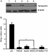
Results: Ferroportin expression in HCC tissue was significantly lower compared to PCLT and normal liver tissue (P <0.05). Moreover, ferroportin expression was related to liver cancer cell de-differentiation, the severity degree in TNM staging, Edmondson-Steiner grading, intrahepatic metastasis and portal vein invasion. In addition, high expression of ferroportin was observed in normal human liver cell lines L02 and HL7702, whereas weak positive expression and even negative expression of ferroportin were observed in HCC cell lines FOCUS, MHCC-97H, HepG2 and SMMC-7721. Furthermore, among the four kinds of HCC cell lines, the expression level of ferroportin was the lowest in MHCC-97H cells.
Conclusions: Ferroportin expression level declines along with the progression of liver cancer, suggesting that the reduction of ferroportin may serve as an important marker for poor HCC prognosis and as a new therapeutic target.
Figures


Similar articles
-
[Expression of mucin 15 in hepatocellular carcinoma and its clinical implications].Nan Fang Yi Ke Da Xue Xue Bao. 2014 Nov;34(11):1611-5. Nan Fang Yi Ke Da Xue Xue Bao. 2014. PMID: 25413059 Chinese.
-
[Over-expression of miR-141 inhibits the proliferation, invasion and migration of hepatocellular carcinoma MHCC-97H cells].Xi Bao Yu Fen Zi Mian Yi Xue Za Zhi. 2016 Aug;32(8):1083-7. Xi Bao Yu Fen Zi Mian Yi Xue Za Zhi. 2016. PMID: 27412940 Chinese.
-
TRIM52 up-regulation in hepatocellular carcinoma cells promotes proliferation, migration and invasion through the ubiquitination of PPM1A.J Exp Clin Cancer Res. 2018 Jun 13;37(1):116. doi: 10.1186/s13046-018-0780-9. J Exp Clin Cancer Res. 2018. PMID: 29898761 Free PMC article.
-
Pathophysiological role of ion channels and transporters in hepatocellular carcinoma.Cancer Gene Ther. 2024 Nov;31(11):1611-1618. doi: 10.1038/s41417-024-00782-8. Epub 2024 Jul 24. Cancer Gene Ther. 2024. PMID: 39048663 Free PMC article. Review.
-
Preclinical human and murine models of hepatocellular carcinoma (HCC).Clin Res Hepatol Gastroenterol. 2024 Aug;48(7):102418. doi: 10.1016/j.clinre.2024.102418. Epub 2024 Jul 14. Clin Res Hepatol Gastroenterol. 2024. PMID: 39004339 Review.
Cited by
-
Downregulation of FPN1 acts as a prognostic biomarker associated with immune infiltration in lung cancer.Aging (Albany NY). 2021 Mar 10;13(6):8737-8761. doi: 10.18632/aging.202685. Epub 2021 Mar 10. Aging (Albany NY). 2021. PMID: 33714956 Free PMC article.
-
Iron-Bound Lipocalin-2 from Tumor-Associated Macrophages Drives Breast Cancer Progression Independent of Ferroportin.Metabolites. 2021 Mar 19;11(3):180. doi: 10.3390/metabo11030180. Metabolites. 2021. PMID: 33808732 Free PMC article.
-
In Situ OH Generation from O2- and H2O2 Plays a Critical Role in Plasma-Induced Cell Death.PLoS One. 2015 Jun 5;10(6):e0128205. doi: 10.1371/journal.pone.0128205. eCollection 2015. PLoS One. 2015. PMID: 26046915 Free PMC article.
-
Regulation of cellular iron metabolism and its implications in lung cancer progression.Med Oncol. 2014 Jul;31(7):28. doi: 10.1007/s12032-014-0028-2. Epub 2014 May 27. Med Oncol. 2014. PMID: 24861923 Review.
-
Retraction note: Ferroportin in the progression and prognosis of hepatocellular carcinoma.Eur J Med Res. 2015 Mar 26;20(1):38. doi: 10.1186/s40001-015-0132-6. Eur J Med Res. 2015. PMID: 25890348 Free PMC article. No abstract available.
References
-
- Okuda K. Hepatocellular carcinoma. J Hepatol. 2000;18:225–237. - PubMed
-
- Schlesinger S, Aleksandrova K, Pischon T, Fedirko V, Jenab M, Trepo E, Boffetta P, Dahm CC, Overvad K, Tjonneland A, Halkjær J, Fagherazzi G, Boutron-Ruault MC, Carbonnel F, Kaaks R, Lukanova A, Boeing H, Trichopoulou A, Bamia C, Lagiou P, Palli D, Grioni S, Panico S, Tumino R, Vineis P, Bueno-de-Mesquita HB, van den Berg S, Peeters PH, Braaten T, Weiderpass E. et al.Abdominal obesity, weight gain during adulthood and risk of liver and biliary tract cancer in a European cohort. Int J Cancer. 2013;18:645–657. doi: 10.1002/ijc.27645. - DOI - PubMed
Publication types
MeSH terms
Substances
LinkOut - more resources
Full Text Sources
Other Literature Sources
Medical
Miscellaneous

