. 2014 Jan 13;28(1):19-29.
doi: 10.1016/j.devcel.2013.11.012. Epub 2013 Dec 19.

Structural insights into assembly and regulation of the plasma membrane phosphatidylinositol 4-kinase complex

Affiliations

Structural insights into assembly and regulation of the plasma membrane phosphatidylinositol 4-kinase complex

Xudong Wu et al. Dev Cell. .

Abstract

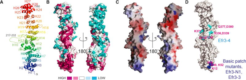
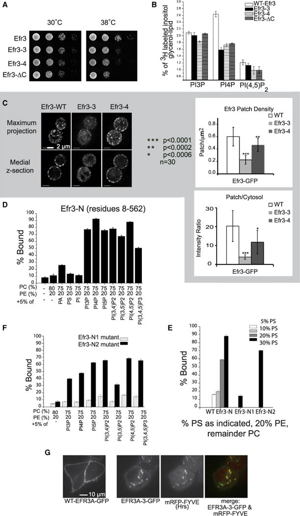
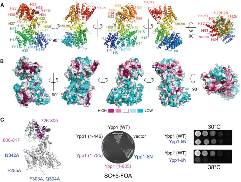
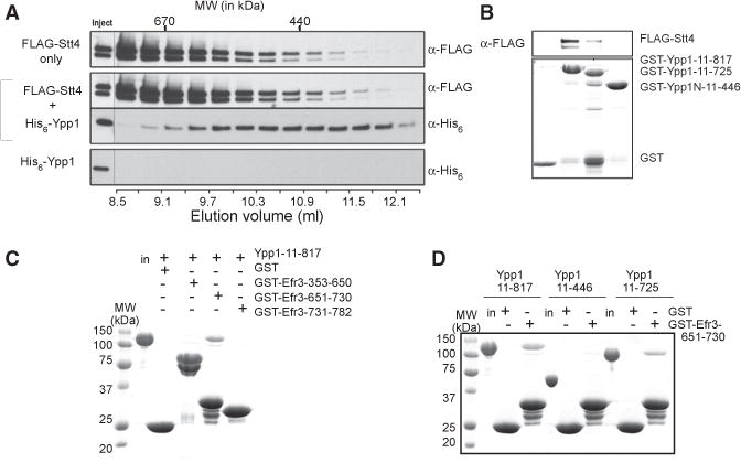
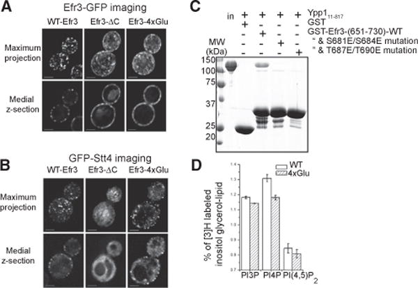
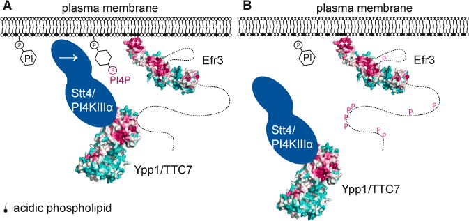
Plasma membrane PI4P helps determine the identity of this membrane and plays a key role in signal transduction as the precursor of PI(4,5)P2 and its metabolites. Here, we report the atomic structure of the protein scaffold that is required for the plasma membrane localization and function of Stt4/PI4KIIIα, the PI 4-kinase responsible for this PI4P pool. Both proteins of the scaffold, Efr3 and YPP1/TTC7, are composed of α-helical repeats, which are arranged into a rod in Efr3 and a superhelix in Ypp1. A conserved basic patch in Efr3, which binds acidic phospholipids, anchors the complex to the plasma membrane. Stt4/PI4KIIIα is recruited by interacting with the Ypp1 C-terminal lobe, which also binds to unstructured regions in the Efr3 C terminus. Phosphorylation of this Efr3 region counteracts Ypp1 binding, thus providing a mechanism through which Stt4/PI4KIIIα recruitment, and thus a metabolic reaction of fundamental importance in cell physiology, can be regulated.


Figures

Figure 1
Figure 1. Structure of Efr3-N
(A) Ribbons diagram, colored blue to red from N terminus to C terminus. (B) Conservation in Efr3-N. Orientation at left is as in (A). Surfaces around the N terminus of Efr3-N and in the middle of the rod are highly conserved. (C) Electrostatic potential. The conserved surface at the N terminus is basic. (D) Location of mutations mapped onto structure. Residues H67 and R69, which are mutated in Efr3-3 and Efr3-N2, are in the conserved basic patch, but not visible here. Recombinant Efr3-3 is well folded as assessed by CD (Figure S2), but Efr3-4 aggregates in vitro.
Figure 2
Figure 2. Conserved Surfaces in Efr3-N Are Important for Function
(A) Yeast strains with Efr3 mutants are growth impaired as compared to those with wild-type protein. (B) Cellular phosphoinositide levels in wild-type and Efr3 mutant strains. PI4P levels are reduced by 30%–40% in the Efr3-3, Efr3-4, and Efr3-ΔC mutants. Error bars are SD. (C) Localization of Efr3-GFP constructs in yeast and quantitation. Membrane localization is reduced for Efr3-3, which has mutations in the conserved basic patch. Student’s t test was used to calculate p values. (D) Liposome binding assays. Experiments shown are sedimentation assays carried out in triplicate; error bars represent SD. Efr3 associates with phosphoinositide-containing liposomes, though nonspecifically. Similar results were obtained using flotation assays (Figure S3). (E) Efr3 also associates with liposomes containing phosphatidylserine, which comprises ~30% of the plasma membrane. Binding is reduced in Efr3-N1 when residues (K12A, R46D, K49A, and K52A) in the conserved basic patch are mutated, but not in Efr3-N2, in which a different and smaller subset of basic patch residues (H67E and R69A) is altered. (F) Binding to phosphoinositide-containing liposomes is significantly reduced for Efr3-N, 1 but not Efr3-N2. (G) Murine EFR3A-GFP and the basic-patch mutant EFR3A-3-GFP were expressed in HeLa cells. EFR3A-3 colocalizes with the endosomal/lysosomal marker mRFP-Fyve and does not go to the plasma membrane like wild-type protein.
Figure 3
Figure 3. Ypp1 Is a HEAT-Repeat Superhelix
(A) Ribbons diagram, colored blue to red from the N terminus to the C terminus. The very C terminus is inserted into the center of the Ypp1 spiral and is marked with *. See also Figure S4. (B) Surface conservation, with orientation as in (A). (C) At left, C-terminal deletions in Ypp1 and residues that were mutated in Ypp1-δN are indicated. In middle, wild-type Ypp1 and Ypp1-δN, but not C-terminally truncated Ypp1 constructs, can rescue Ypp1 null yeast strains. At right, Ypp1-δN has a growth defect at restricted temperature. (See Figure S2 for CD profiles of Ypp1 mutants.)
Figure 4
Figure 4. The C-Terminal Portion of Ypp1 Is Required for Interactions with Stt4 and Efr3
(A) Gel filtration chromatography (Superdex200 10/300) showing elution profiles of affinity-purified FLAG-Stt4 (top panel), His6-Ypp1 (bottom panel), and a mixture of FLAG-Stt4 and His6-Ypp1 (middle panels). Stt4 is partially aggregated and mostly in the void volume, with only a small monodisperse fraction. Ypp1 alone elutes at 13.75 ml. Its elution peak shifts in the presence of Stt4, and Ypp1 comigrates with monodisperse Stt4. They elute at 10.8 ml, similarly to the ~440 kDa ferritin calibration standard. The column was calibrated with HMW calibration standards (Biorad) and ferritin (Sigma). (B) Pull-down of recombinant FLAG-tagged Stt4 by purified Ypp1 constructs. Stt4 interacts with fragments that include C-terminal portions of Ypp1 and not with the N-terminal half of Ypp1. (C) Pull-down of Ypp1 by C-terminal fragments of Efr3. Ypp1 interacts with an Efr3 fragment including residues 651–730. (D) Pull-down of Ypp1 constructs by an Efr3 C-terminal fragment. Only fragments that include C-terminal portions of Ypp1 interact with Efr3; Ypp1-N does not.
Figure 5
Figure 5. Efr3 C-Terminal Phosphorylation Modulates the Efr3-Ypp1 Interaction and Stt4 Complex Assembly
(A) Efr3-ΔC-GFP localizes to the plasma membrane in yeast, but does not form punctae like wild-type Efr3-GFP. The Efr3-4×Glu mutant has a similar phenotype. (B) GFP-Stt4 is mostly cytosolic when expressed in a Efr3-ΔC background and does not localize to the plasma membrane. Stt4-GFP plasma membrane localization is also reduced in the Efr3-4×Glu background. (C) Ypp1 pull-down with C-terminal fragments of Efr3 (residues 651–730). Mutating S681/S684 or T687/T690 to glutamate to mimic their phosphorylation reduced the interaction between Ypp1 and Efr3. (D) Cellular PI4P levels are reduced by ~10% in yeast strains with Efr3-4×Glu as compared to wild-type protein.
Figure 6
Figure 6. Stt4/PI4KIIIα Targeting to the Plasma Membrane
(A) A conserved basic patch at the N-terminal end of Efr3 mediates complex association with the plasma membrane. The Efr3 C terminus and Stt4/PI4KIIIα both interact with the C-terminal portion of Ypp1. As yet, no function has been assigned to conserved surfaces at the middle of Efr3-N or in the Ypp1 N-terminal half. (B) Stt4/PI4KIIIα recruitment to the plasma membrane is reduced when the Efr3 C terminus is phosphorylated to interrupt the Efr3-Ypp1 interaction. P indicates that the Efr3 C terminus is heavily phosphorylated in vivo.

References

    1. Abramoff MD, Magalhães PJ, Ram SJ. Image processing with ImageJ. Biophotonics International. 2004;11:36–42.
    1. Adams PD, Grosse-Kunstleve RW, Hung LW, Ioerger TR, McCoy AJ, Moriarty NW, Read RJ, Sacchettini JC, Sauter NK, Terwilliger TC. PHENIX: building new software for automated crystallographic structure determination. Acta Crystallogr D Biol Crystallogr. 2002;58:1948–1954. - PubMed
    1. Altan-Bonnet N, Balla T. Phosphatidylinositol 4-kinases: hostages harnessed to build panviral replication platforms. Trends Biochem Sci. 2012;37:293–302. - PMC - PubMed
    1. Ashkenazy H, Erez E, Martz E, Pupko T, Ben-Tal N. ConSurf 2010: calculating evolutionary conservation in sequence and structure of proteins and nucleic acids. Nucleic Acids Res. 2010;38:W529–W533. Web Server issue. - PMC - PubMed
    1. Audhya A, Emr SD. Stt4 PI 4-kinase localizes to the plasma membrane and functions in the Pkc1-mediated MAP kinase cascade. Dev Cell. 2002;2:593–605. - PubMed

MeSH terms

Substances