. 2014 Jan 2;94(1):120-8.
doi: 10.1016/j.ajhg.2013.11.020. Epub 2013 Dec 19.

A noncoding expansion in EIF4A3 causes Richieri-Costa-Pereira syndrome, a craniofacial disorder associated with limb defects

Affiliations

A noncoding expansion in EIF4A3 causes Richieri-Costa-Pereira syndrome, a craniofacial disorder associated with limb defects

Francine P Favaro et al. Am J Hum Genet. .

Abstract

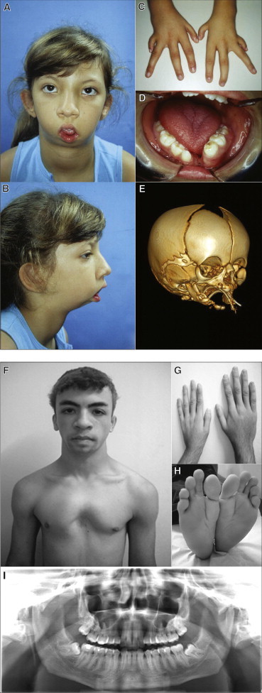
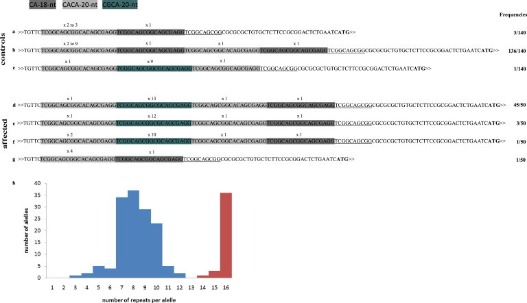
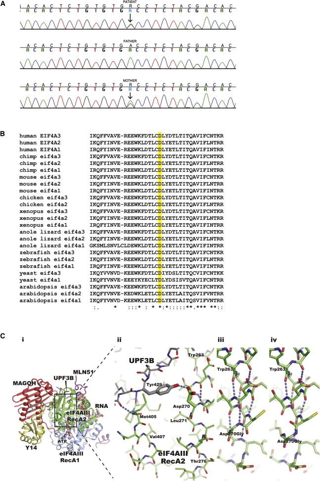
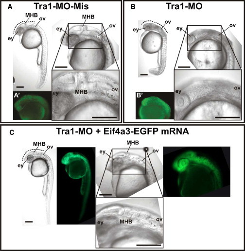
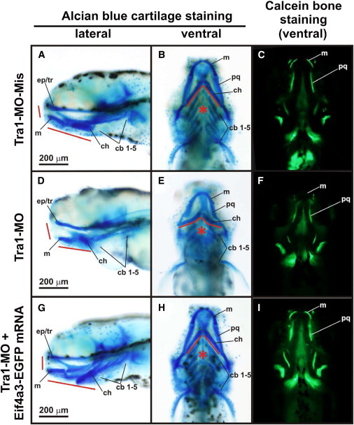
Richieri-Costa-Pereira syndrome is an autosomal-recessive acrofacial dysostosis characterized by mandibular median cleft associated with other craniofacial anomalies and severe limb defects. Learning and language disabilities are also prevalent. We mapped the mutated gene to a 122 kb region at 17q25.3 through identity-by-descent analysis in 17 genealogies. Sequencing strategies identified an expansion of a region with several repeats of 18- or 20-nucleotide motifs in the 5' untranslated region (5' UTR) of EIF4A3, which contained from 14 to 16 repeats in the affected individuals and from 3 to 12 repeats in 520 healthy individuals. A missense substitution of a highly conserved residue likely to affect the interaction of eIF4AIII with the UPF3B subunit of the exon junction complex in trans with an expanded allele was found in an unrelated individual with an atypical presentation, thus expanding mutational mechanisms and phenotypic diversity of RCPS. EIF4A3 transcript abundance was reduced in both white blood cells and mesenchymal cells of RCPS-affected individuals as compared to controls. Notably, targeting the orthologous eif4a3 in zebrafish led to underdevelopment of several craniofacial cartilage and bone structures, in agreement with the craniofacial alterations seen in RCPS. Our data thus suggest that RCPS is caused by mutations in EIF4A3 and show that EIF4A3, a gene involved in RNA metabolism, plays a role in mandible, laryngeal, and limb morphogenesis.


Figures

Figure 1
Figure 1
Clinical and Radiographic Aspects of Typical and Milder Forms of RCPS (A–D) Affected individual 2 at 9 years of age, illustrating (A, B) typical facial features of the syndrome including micrognathia and microstomia, (C) hypoplasia of fingers and clinodactyly, and (D) absence of lower central incisors and median mandibular cleft. (E) CT scan of the skull of affected individual 23 at 12 days of age. Note the very rudimentary mandibular formation with large medial cleft, micrognathia, and incomplete zygomatic arches. (F–I) Affected individual 25, illustrating (F) absence of microstomia and presence of pectus excavatum, (G) short left hand with hypoplastic thumbs, (H) feet with characteristically abnormal shape, and (I) normal fusion of mandible.
Figure 2
Figure 2
Representation of the 5′ UTR of EIF4A3 Alleles According to the Pattern of the Repeats and Their Distribution Frequency (A–C) Three patterns of repeats in control alleles: note that the number of CA-18-nt motifs varied among the different allele patterns and that the CGCA-20-nt motif was present in only 1 out of 140 alleles. (D–F) Three patters of repeats in affected alleles: the 15- and 16-repeat alleles have the same underlying structure (D, E), whereas the 14-repeat allele contains an additional copy of the CACA-20-nt motif (F). (G) The nonexpanded 5′ UTR in cis with the nucleotide substitution c.809A>G (p.Asp270Gly) allele in individual 25 corresponds to one of the patterns of the control individuals (A). The first thymidine (T) at the 5′ end corresponds to position +38 of the transcription initiation site and the ATG corresponds to the translation initiation codon. Underlined sequence downstream of the last 18-nt motif represents a partially conserved motif in all alleles. (H) Distribution of the alleles per repeat size. Control sample alleles are shown in blue and affected alleles shown in red. The counting of the alleles in affected individuals took into consideration relatedness.
Figure 3
Figure 3
Description of the Missense Mutation in EIF4A3, Evolutionary Comparative Analysis, and Structural Analysis (A) Sequence analysis of exon 8 showing the A to G substitution at position c.809 (indicated by an arrow), leading to the amino acid substitution p.Asp270Gly. The online tool PolyPhen-2 predicts this mutation as possibly damaging with a score of 0.860 (sensitivity: 0.72 and specificity: 0.89). (B) Comparative sequence analysis of EIF4A3 encoded orthologous proteins and its paralogs, showing that the Asp270 (highlighted D) is highly conserved throughout evolution. (C) Structure of the core human exon junction complex bound to a C-terminal fragment of UPF3B (PDB entry 2XB2 for RCSB). (i) Overall view: the RNA and ATP binding eIF4A3 is shown with its RecA1 and RecA2 domains colored blue and green, respectively. A fragment of MLN51 (magenta) encloses both RecA domains and also forms part of the binding site for the 5′ end of the RNA. MAGOH (red) contacts both domains of eIF4AIII, the MLN51 fragments, and positions Y14 (yellow) in the complex. The C-terminal UPF3B fragment (gray) interacts with both Y14 and the eIF4AIII RecA2 domain. (ii) Close-up on the UPF3B Tyr429 interaction with the Asp270 in the eIF4A3 RecA2 domain. Selected putative hydrogen bonds of relevance for the Asp-Gly mutation are indicated with blue dashed lines. (iii) Selected model of the glycine mutant with a secondary structure similar to that of the experimental structures of wild-type eIF4AIII. (iv) Selected model of the glycine mutant with a disrupted secondary structure compared to the experimental structures of wild-type eIF4AIII. Models were prepared with Modeler.
Figure 4
Figure 4
Zebrafish eif4a3 Knockdown via Morpholinos Microinjected zebrafish embryos staged at 24 hr postfertilization (hpf) presented an observable phenotype. (A) Control embryo microinjected with mispaired MO (TRA1-MO-Mis). (B) Morphant embryo microinjected with MO designed to block eif4a3-mRNA translation (TRA1-MO). (C) Rescued embryo microinjected with TRA1-MO and mRNA coding for eif4a3 fused to EGFP (eif4a3-EGFP mRNA). Lateral views of embryos were registered under stereoscopic microscope (whole body at the upper left of each panel) or with two different magnifications under differential interference contrast microscope (anterior-most region at the upper and lower right of each panel). Alterations in the morphants’ trunk were observed but not reproducible and only phenotyping at the craniofacial level was done. Dotted lines mark regions between eyes (ey) and otic vesicle (ov), which include the midbrain-hindbrain border (MHB) and showed darkened tissue in morphants, suggesting the presence of apoptotic cells. Lateral views of 24 hpf embryos after acridine orange staining, by standardized protocols, are shown in (A′) and (B′). Green fluorescence panels in (C) show expression of eif4a3-EGFP in rescued embryo. Scale bars represent 200 μm. TRA1-MO sequence: TGTGACGGATTTCGGTGTAAATTAC. TRA1-MO-Mis sequence: TGTCACCGATTTCCGTCTAAAATAC.
Figure 5
Figure 5
Craniofacial Phenotype in Zebrafish eif4a3 Morphant Larvae Five days postfertilization (dpf) larvae microinjected with TRA1-MO-Mis (A–C), TRA1-MO (D–F), and TRA1-MO + eif4a3-EGFP mRNA (G–I) were stained with alcian blue to observe cartilage structures (A, D, G, lateral views; B, E, H, ventral views) or with calcein to observe bone structures (C, F, I, ventral views). Eyes were removed to register alcian blue-stained larvae. Craniofacial precursors in morphant larvae were severely affected showing hypoplasia of numerous craniofacial cartilages (red lines), which is evident in the rostral and jaw elements. Impairment of pharyngeal arches development was also observed (asterisks). Abbreviations are as follows: cb1–5, ceratobranchial arches 1–5; ch, ceratohyal; ep/tr, ethmoid plate/trabecula; m, Meckel’s cartilage; pq, palatoquadrate. Scale bars represent 200 μm.

References

    1. Favaro F.P., Zechi-Ceide R.M., Alvarez C.W., Maximino L.P., Antunes L.F.B.B., Richieri-Costa A., Guion-Almeida M.L. Richieri-Costa-Pereira syndrome: a unique acrofacial dysostosis type. An overview of the Brazilian cases. Am. J. Med. Genet. A. 2011;155A:322–331. - PubMed
    1. Ferreira de Lima R.L., Moretti-Ferreira D., Richieri-Costa A., Murray J.C. Identity by descent and candidate gene mapping of Richieri-Costa and Pereira syndrome. Am. J. Med. Genet. A. 2003;122A:56–58. - PubMed
    1. Abecasis G.R., Cherny S.S., Cookson W.O., Cardon L.R. Merlin—rapid analysis of dense genetic maps using sparse gene flow trees. Nat. Genet. 2002;30:97–101. - PubMed
    1. Jordan C.T., Cao L., Roberson E.D., Duan S., Helms C.A., Nair R.P., Duffin K.C., Stuart P.E., Goldgar D., Hayashi G. Rare and common variants in CARD14, encoding an epidermal regulator of NF-kappaB, in psoriasis. Am. J. Hum. Genet. 2012;90:796–808. - PMC - PubMed
    1. Becker-Heck A., Zohn I.E., Okabe N., Pollock A., Lenhart K.B., Sullivan-Brown J., McSheene J., Loges N.T., Olbrich H., Haeffner K. The coiled-coil domain containing protein CCDC40 is essential for motile cilia function and left-right axis formation. Nat. Genet. 2011;43:79–84. - PMC - PubMed

Publication types

MeSH terms

Substances

Supplementary concepts