. 2014 Feb 6;14(2):188-202.
doi: 10.1016/j.stem.2013.12.001. Epub 2013 Dec 19.

In vivo direct reprogramming of reactive glial cells into functional neurons after brain injury and in an Alzheimer's disease model

Affiliations

In vivo direct reprogramming of reactive glial cells into functional neurons after brain injury and in an Alzheimer's disease model

Ziyuan Guo et al. Cell Stem Cell. .

Abstract

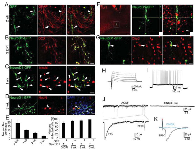
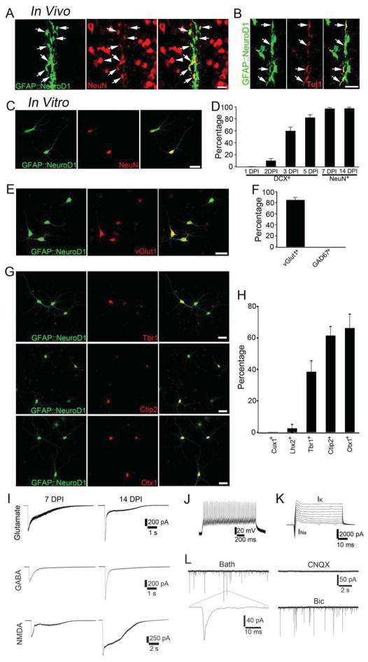
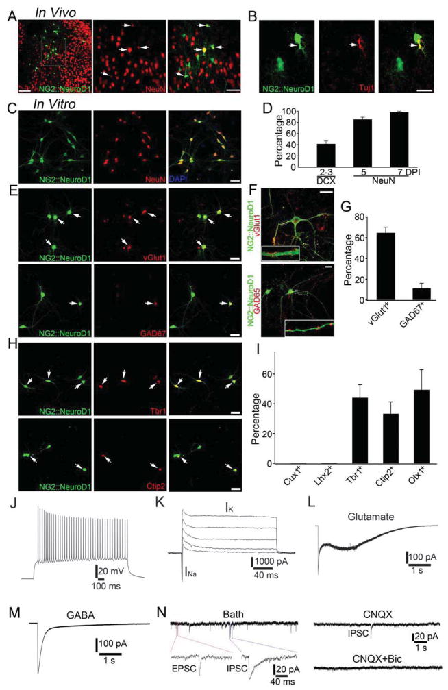
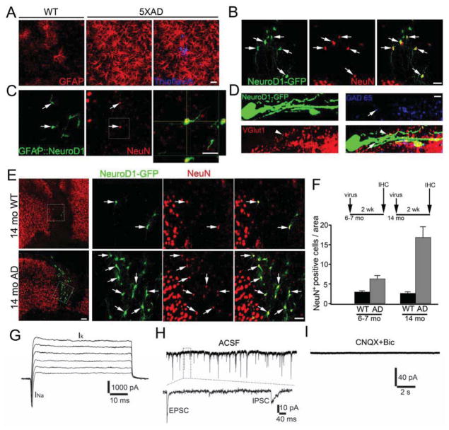
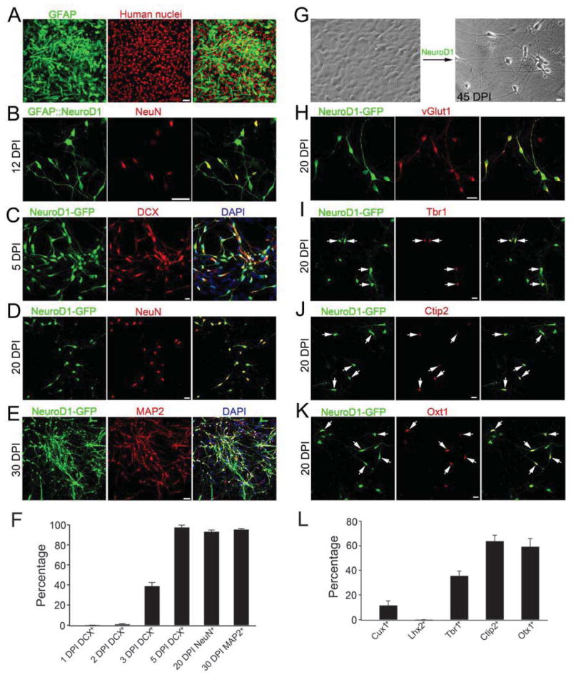
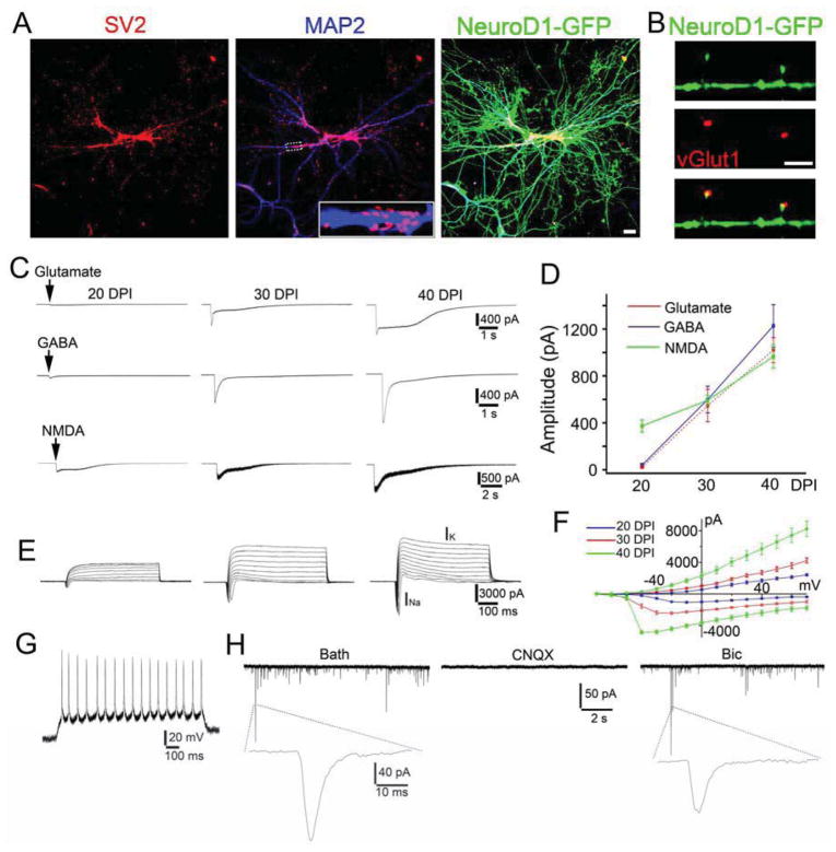
Loss of neurons after brain injury and in neurodegenerative disease is often accompanied by reactive gliosis and scarring, which are difficult to reverse with existing treatment approaches. Here, we show that reactive glial cells in the cortex of stab-injured or Alzheimer's disease (AD) model mice can be directly reprogrammed into functional neurons in vivo using retroviral expression of a single neural transcription factor, NeuroD1. Following expression of NeuroD1, astrocytes were reprogrammed into glutamatergic neurons, while NG2 cells were reprogrammed into glutamatergic and GABAergic neurons. Cortical slice recordings revealed both spontaneous and evoked synaptic responses in NeuroD1-converted neurons, suggesting that they integrated into local neural circuits. NeuroD1 expression was also able to reprogram cultured human cortical astrocytes into functional neurons. Our studies therefore suggest that direct reprogramming of reactive glial cells into functional neurons in vivo could provide an alternative approach for repair of injured or diseased brain.


Figures

Figure 1
Figure 1. In vivo conversion of reactive glial cells into functional neurons after brain injury
(A) Injecting control retrovirus expressing GFP (green) into mouse cortex revealed GFAP-positive reactive astrocytes (red) in the injury site (14 days post injection, DPI). (B–C) NeuroD1-IRES-GFP infected cells (green) were immunopositive for neuronal markers DCX (B, 3 DPI) and NeuN (C, 7 DPI). Note a significant number of NeuN-positive neurons in the injury site after NeuroD1 infection. (D) After 21 DPI, NeuroD1-converted neurons (NeuN-positive, arrowhead) showed extensive neurites. Scale bar, 20 μm for (A) and (D); 40 μm for (B) and (C). (E) Quantified data showing the number of converted neurons per imaged area (40x, 0.1 mm2) and conversion efficiency after NeuroD1 infection. (F–G) NeuroD1-converted neurons were immunopositive for cortical neuron marker Tbr1 (F) and deep layer marker Ctip2 (G, 12 DPI). Scale bars: 100 μm for low power image, 40 μm for high power image. (H–I) Representative traces from cortical slice recordings showing Na+ and K+ currents (H) and repetitive action potentials (I) in NeuroD1-converted neurons (30 DPI). (J) Representative traces showing spontaneous synaptic events in a NeuroD1-converted neuron (26 DPI) in cortical slice recording (CNQX, 10 μM; Bic, 20 μM). (K) Evoked synaptic events recorded from a converted neuron. See also Suppl. Fig. 1–3.
Figure 2
Figure 2. NeuroD1 converts astrocytes into glutamatergic neurons
(A–B) In vivo injection of GFAP promoter-driven NeuroD1-IRES-GFP (green) retrovirus revealed astrocyte-converted neurons immunopositive for NeuN (A) and Tuj1 (B). (C) Cultured mouse cortical astrocytes were converted into NeuN-positive neurons. (D) Time course of GFAP::NeuroD1 conversion efficiency after infecting cultured mouse astrocytes. (E–F) Astrocyte-converted neurons were positive for VGluT1 (E) but negative for GAD67 (F). (G–H) Immunostaining with cortical layer neuronal markers showed deep layer neuronal properties (Ctip2 and Otx1) after NeuroD1-induced conversion. Scale bars: 20 μm for (A–C) and (E), and 40 μm for (G). (I) Mouse astrocyte-converted neurons showed large glutamate, GABA, and NMDA receptor currents within 2 weeks after NeuroD1 infection. Average GABA current, 7 DPI, 405 ± 97 pA, n=8; 14 DPI, 861 ± 55 pA, n=13. Average glutamate current, 7 DPI, 517 ± 145 pA, n=7; 14 DPI, 1060 ± 159 pA, n=9. Average NMDA current, 7 DPI, 676 ± 118 pA, n=7; 14 DPI, 1315 ± 95, n=7. (J–K) Mouse astrocyte-converted neurons showed repetitive action potentials (J) and large INa and IK (K). (L) Spontaneous synaptic events recorded from mouse astrocyte-converted neurons. All events were blocked by CNQX but not Bic, suggesting that they were glutamatergic events. Also see Suppl. Fig. 4.
Figure 3
Figure 3. NeuroD1 converts NG2 cells into glutamatergic and GABAergic neurons
(A–B) In vivo injection of NG2::NeuroD1-GFP retrovirus revealed the conversion of NG2 cells into neuronal cells positive for NeuN (A) or Tuj1(B) (8 DPI). (C–D) Cultured NG2 cells were converted into NeuN-positive neurons within one week after infection by NG2::NeuroD1. (E–G) NG2 cell-converted neurons after NeuroD1-infection were immunopositive for both VGluT1 (>60%) and GAD67 (10%). VGluT1 and GAD65 immunostaining also showed glutamatergic and GABAergic puncta on converted neural dendrites (F). (H–I) Cortical layer neuronal marker immunostaining showed deep layer neuronal properties (Ctip2 and Otx1) after NeuroD1-induced conversion of NG2 cells. Scale bars: 40 μm for (A), (C), (E), and (H); 20 μm for (B) and (F). (J–K) NG2-converted neurons showed repetitive action potentials (J; n = 9) and large sodium and potassium currents (K; n = 10). (L–M) NG2-converted neurons showed large glutamate-evoked current (L; n = 7) and GABA-evoked current (M; n = 7). (N) Spontaneous synaptic events recorded from NG2-converted neurons showed both glutamatergic and GABAergic events, confirming that NeuroD1 can convert NG2 cells into both excitatory and inhibitory neurons.
Figure 4
Figure 4. NeuroD1 converts reactive glial cells into functional neurons in AD mouse brain in vivo
(A) Reactive astrocytes (labeled by GFAP, red) in 5xFAD mouse cortex (5-month old) were significantly increased compared to that in WT cortex. Aβ plaques were labeled by thioflavin-S (blue). (B) NeuroD1-infected cells (16 DPI) in AD mouse cortex (7-month old) showed clear neuron-like morphology (green) and NeuN staining (red). (C) Injecting GFAP::NeuroD1 retrovirus into AD cortex also converted astrocytes into NeuN-positive neurons (7 DPI). (D) NeuroD1-converted neurons in the AD brain were innervated by glutamatergic (VGluT1, red) and GABAergic terminals (GAD65, blue). Scale bars: 20 μm for (A) and (C); 40 μm for (B); 5 μm for (D). (E) Efficient induction of many new neurons in 14-month old AD animals after NeuroD1-GFP retroviral infection. Scale bar: 100 μm for low power image, 40 μm for high power image. (F) Quantified data showing enhanced neural conversion in AD animals compared to WT animals, likely due to more reactive glial cells in old AD brain. (G) Representative traces of sodium and potassium currents recorded from NeuroD1-infected cells in AD cortical slices. (H) Spontaneous synaptic events recorded from NeuroD1-converted neurons (28 DPI) in AD cortical slices. (I) All synaptic events were blocked by CNQX (10 μM) and BIC (20 μM).
Figure 5
Figure 5. Conversion of cultured human astrocytes into functional neurons
(A) The majority of cultured human astrocytes were labeled by GFAP (green). (B) Infection by GFAP::NeuroD1 retrovirus converted human astrocytes into NeuN-positive neurons. (C–E) NeuroD1-induced conversion of human astrocytes into neurons as shown by a series of neuronal markers DCX (C), NeuN (D), and MAP2 (E). (F) Quantified data showing a significant increase of conversion efficiency during 3 – 5 DPI. (G) Phase contrast images showing NeuroD1-induced morphological change from astrocytes (left) to neurons (right, 45 DPI). (H) Human astrocyte-converted neurons were immunopositive for VGluT1. (I–K) Cortical layer neuronal markers revealed that human astrocyte-converted neurons were immunopositive for Tbr1 (I), Ctip2 (J), and Otx1 (K). (L) Quantitative analysis of human astrocyte-converted neurons labeled by superficial (Cux1 and Lhx2) or deep layer (Ctip2 and Otx1) neuronal markers. Scale bars: 50 μm for (A) and (E); 20 μm for panels (C–D) and (G–K); 40 μm for panel (B). See also Suppl. Fig. 5–6.
Figure 6
Figure 6. Functional characterization of human astrocyte-converted neurons
(A) Synaptic puncta (SV2, red) on the dendrites (MAP2, blue) of human astrocyte-converted neurons (green, 45 DPI) after NeuroD1 infection. (B) High power image showing VGlut1 puncta (red) co-localized with dendritic spines on NeuroD1-converted neurons. Scale bars: 20 μm for panel (A); 10 μm for panel (B). (C–D) Representative traces (C) and quantitative analysis (D) of the receptor currents induced by bath application of glutamate (100 μM), GABA (100 μM), and NMDA (100 μM). (E–F) Representative traces of Na+ and K+ currents (E) and their I–V curve (F) recorded from NeuroD1-converted neurons. (G) Representative trace of repetitive action potentials in NeuroD1-converted neurons (20 DPI). (H) Representative traces of spontaneous synaptic events in NeuroD1-converted human neurons (40 DPI). Note that all synaptic events were blocked by CNQX (10 μM) but not by Bic (20 μM), suggesting that human astrocyte-converted neurons induced by NeuroD1 expression were glutamatergic neurons.

Comment in

References

    1. Aguzzi A, Barres BA, Bennett ML. Microglia: scapegoat, saboteur, or something else? Science. 2013;339:156–161. - PMC - PubMed
    1. Ambasudhan R, Talantova M, Coleman R, Yuan X, Zhu S, Lipton SA, Ding S. Direct reprogramming of adult human fibroblasts to functional neurons under defined conditions. Cell Stem Cell. 2011;9:113–118. - PMC - PubMed
    1. Bayart E, Cohen-Haguenauer O. Technological overview of iPS induction from human adult somatic cells. Current gene therapy. 2013;13:73–92. - PMC - PubMed
    1. Bergles DE, Jabs R, Steinhauser C. Neuron-glia synapses in the brain. Brain research reviews. 2010;63:130–137. - PMC - PubMed
    1. Boutin C, Hardt O, de Chevigny A, Core N, Goebbels S, Seidenfaden R, Bosio A, Cremer H. NeuroD1 induces terminal neuronal differentiation in olfactory neurogenesis. Proc Natl Acad Sci U S A. 2010;107:1201–1206. - PMC - PubMed

Publication types

MeSH terms