. 2014 Jan 6;24(1):19-28.
doi: 10.1016/j.cub.2013.11.029. Epub 2013 Dec 19.

Autophagy in oncogenic K-Ras promotes basal extrusion of epithelial cells by degrading S1P

Affiliations

Autophagy in oncogenic K-Ras promotes basal extrusion of epithelial cells by degrading S1P

Gloria Slattum et al. Curr Biol. .

Abstract

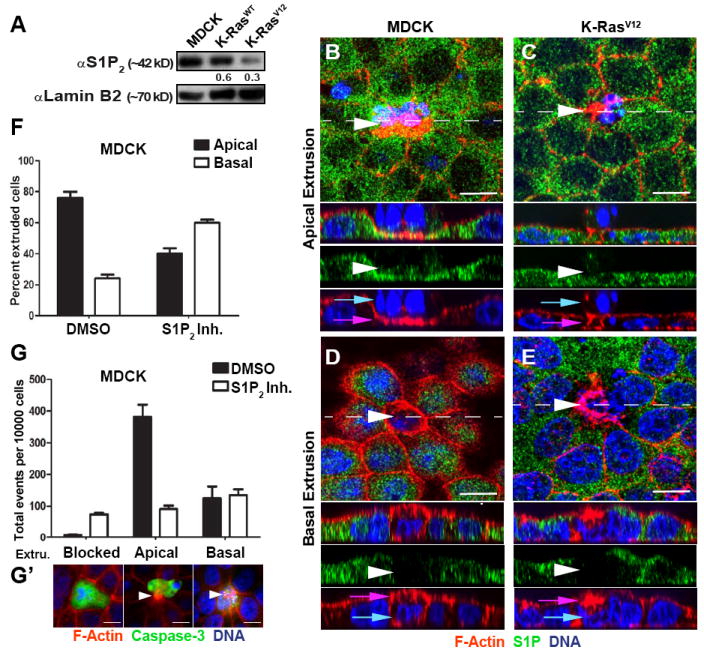
Background: To maintain a protective barrier, epithelia extrude cells destined to die by contracting a band of actin and myosin. Although extrusion can remove cells triggered to die by apoptotic stimuli, to maintain constant cell numbers, epithelia extrude live cells, which later die by anoikis. Because transformed cells may override anoikis and survive after extrusion, the direction of extrusion has important consequences for the extruded cell's fate. As most cells extrude apically, they are typically eliminated through the lumen; however, cells with upregulated survival signals that extrude basally could potentially invade the underlying tissue and migrate to other sites in the body.

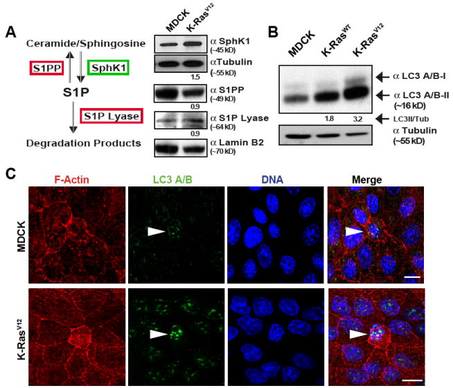
Results: We found that oncogenic K-Ras cells predominantly extrude basally, rather than apically, in a cell-autonomous manner and can survive and proliferate after extrusion. Expression of K-Ras(V12) downregulates the bioactive lipid sphingosine 1-phosphate (S1P) and its receptor S1P2, both of which are required for apical extrusion. Surprisingly, the S1P biosynthetic pathway is not affected because the S1P precursor, sphingosine kinase, and the degradative enzymes S1P lyase and S1PP phosphatase are not significantly altered. Instead, we found that high levels of autophagy in extruding Ras(V12) cells leads to S1P degradation. Disruption of autophagy chemically or genetically in K-Ras(V12) cells rescues S1P localization and apical extrusion.

Conclusions: Oncogenic K-Ras cells downregulate both S1P and its receptor S1P2 to promote basal extrusion. Because live basally extruding cells can survive and proliferate after extrusion, we propose that basal cell extrusion provides a novel mechanism for cells to exit the epithelium and initiate invasion into the surrounding tissues.


Figures

Figure 1
Figure 1
Epithelial cells expressing K-RasV12 extrude basally in a cell-autonomous manner. Confocal projections and XZ cross-sections (below) showing extruding cells (white arrowheads) (A-C). Control MDCK cells (A) and those expressing YFP-K-RasWT (B) extrude apically (DNA of extruding cell is out of plane with neighboring cells), while those expressing GFP-K-RasV12 (C) extrude basally (DNA of extruding cell is in same plane as neighboring cells). Quantification of cell extrusion events from 5 independent experiments with >1000 extruding cells per cell line (D). MDCK GFP-K-RasV12 cells mixed with wild type MDCK cells 1:100 indicate that K-RasV12 cells (green arrow) drive basal extrusion cell autonomously, where arrowhead indicates basally extruded K-RasV12 cell and asterisks show wild type cells (E). Pink and blue arrows indicate actin ring and DNA, respectively, and dashed line where XZ section was taken. Quantification of 3 independent experiments, where n=600 extruding cells (F). Scale Bar=10 μm; error bars= SEM; P<0.001 by student’s t test comparing control to RasV12.
Figure 2
Figure 2
K-RasV12 does not affect direction of extrusion by altering microtubules dynamics or cell intrinsic polarity. Confocal projections and XZ cross-sections (below) of control MDCK monolayers apically extruding (A-C) and K-RasV12 monolayers basally extruding (D-F). White Arrows point to the microtubules pointing basally during apical extrusion (A) and apically during basal extrusion (D), where asterisks indicate extruding cells location. White arrows show the apical marker ZO-1 (B&E) and the basal marker β-catenin (E&F) are both localized correctly. White arrowhead indicates extruding cell, pink and blue arrows indicate actin ring and DNA, respectively, and dashed line where XZ section was taken. Scale bar=10 μm.
Figure 3
Figure 3
Sphingosine-1-Phosphate (S1P) and the S1P receptor 2 (S1P2) signals apical but not basal extrusion and is misregulated in K-RasV12 expressing cells. (A) Immunoblot analysis comparing the expression levels of S1P2 in control MDCK cells, and MDCK cells expressing K-RasWT or K-RasV12 where lamin B2 serves as a loading control and quantified ratio compared to control is below. Confocal projections and XZ cross-sections (below) showing an apically extruding control MDCK produces high S1P levels (B) whereas an apically extruding K-RasV12 cell does not (C). Basally extruding control (D) and K-RasV12 (E) MDCKs do not produce any S1P. White arrowheads point to dying, extruding cells at live-dead cell interfaces, pink arrows indicate actin ring, blue arrows show DNA, and dashed line where XZ section was taken. Scale Bar=10 μm. (F&G) Apical extrusion is disrupted in control MDCK cells treated with S1P2 antagonist (JTE-013), where (F) shows comparative rates, (G) shows total rates from 3 independent experiments analyzing 300 extrusion events for each experiment; error bars=SEM. P<0.001 by student’s t test comparing control versus each treatment. (G’) shows examples of blocked extrusion, scored by the presence of a faint actin ring that does not contract around a late staged capase-3-positive dying cell, apical extrusion, and basal extrusion.
Figure 4
Figure 4
K-RasV12 extruding cells express high levels of the autophagy marker LC3 A/B. (A) SIP synthetic pathway and immuno-blots comparing the expression levels of key regulators of S1P in control versus K-RasV12 cell lysates where tubulin and lamin serve as loading controls. (B) LC3A/B immunoblots of MDCK, K-RasWT, and K-RasV12 cells, where α-tubulin serves as a loading control, LC3 A/B-I is the cytosolic form whereas LC3 A/B-II is associated with autophagosomes. Ratios compared to the control are below. (C) Confocal projections of control MDCKs and K-RasV12 basal extruding cell, white arrowheads show increased LC3 puncta in extruding cell. Scale Bar=10 μm.
Figure 5
Figure 5
Blocking autophagy in K-RasV12 cells rescues S1P localization and apical extrusion. Confocal section and XZ cross sections (below) of a K-RasV12 monolayer that extrudes primarily basally and lacks SIP (A) but extrudes apically and accumulates S1P puncta with 30μM Chloroquine, which blocks autophagy (B), where white arrowheads indicate extruding cells at the live-dead cell interfaces, pink arrows indicate actin ring, blue arrows show DNA, and dashed line the XZ section. Scale Bar= 10 μm. (C) Quantifications of cell extrusion direction with three autophagy inhibitors from four independent experiments where >1000 extrusions were analyzed per treatment; error bars= SEM; (D) immunoblots show siRNA-mediated knockdown of Atg7 and LC3 II reduction and p62 upregulation. Ratios compared to the control are below. Atg7 knockdown also rescues apical extrusion and S1P (E and F) quantification from <500 total extrusion events from 3 siRNAs experiments. P<0.001 by student’s t test comparing control versus each treatment. (G) Model for how apical extrusion is misregulated in K-RasV12 expressing cells: S1P normally binds S1P2 in surrounding cells to contract basally and squeeze the cell out. However, in K-RasV12 extruding cells S1P is targeted for degradation by increased autophagy. Without S1P, basolateral contraction in neighboring cells is not activated and instead only apical contraction of the dying cell occurs, driving extrusion basally. S1P (blue), autophagosome (yellow), lysosome (green), extruding cell actomyosin contraction (red), neighboring cell actomyosin contraction (maroon).
Figure 6
Figure 6
Basally extruded K-RasV12 cells survive and proliferate in 3D cultures. Cysts from MDCK and K-RasV12-MDCK cells cultured in matrigel for three days. Confocal sections showing that wild type MDCK cysts have clear lumens (A) whereas MDCK K-RasV12 cysts extrude live cells basally (B). Yellow arrowhead, filled lumen and red arrows, live basal extruded cells. Scale bar=20 μm. (C) Quantification of phenotypes over time from 3 independent experiments, n>100 cysts per experiment; error bars= SEM; P<0.001 by student’s t test comparing control to RasV12. Stills from movies of cysts (in Movie S5) filmed at day 3 for 24 hours without UV treatment (D&E). A control MDCK cystb extrudes cells apically into the lumen (yellow arrow) (D), whereas, a K-RasV12 cyst extrudes cells basally into the matrix, which later proliferate (E) (red arrows). Stills from movie of a K-RasV12 cyst UV treated expressing Myosin Light Chain (MLC)-RFP (in Movie S6) show that basal extrusion of a cell occurs by contraction of a myosin ring (F). Time:h:m

References

    1. Rosenblatt J, Raff MC, Cramer LP. An epithelial cell destined for apoptosis signals its neighbors to extrude it by an actin- and myosin-dependent mechanism. Curr Biol. 2001;11:1847–1857. - PubMed
    1. Gu Y, Forostyan T, Sabbadini R, Rosenblatt J. Epithelial cell extrusion requires the sphingosine-1-phosphate receptor 2 pathway. J Cell Biol. 2011;193:667–676. - PMC - PubMed
    1. Eisenhoffer GT, Loftus PD, Yoshigi M, Otsuna H, Chien CB, Morcos PA, Rosenblatt J. Crowding induces live cell extrusion to maintain homeostatic cell numbers in epithelia. Nature. 2012;484:546–549. - PMC - PubMed
    1. Marinari E, Mehonic A, Curran S, Gale J, Duke T, Baum B. Live-cell delamination counterbalances epithelial growth to limit tissue overcrowding. Nature. 2012;484:542–545. - PubMed
    1. Frisch SM, Vuori K, Ruoslahti E, Chan-Hui PY. Control of adhesion-dependent cell survival by focal adhesion kinase. J Cell Biol. 1996;134:793–799. - PMC - PubMed

Publication types