. 2014 Jan 8;81(1):179-94.
doi: 10.1016/j.neuron.2013.10.020. Epub 2013 Dec 19.

Flow of cortical activity underlying a tactile decision in mice

Affiliations

Flow of cortical activity underlying a tactile decision in mice

Zengcai V Guo et al. Neuron. .

Abstract

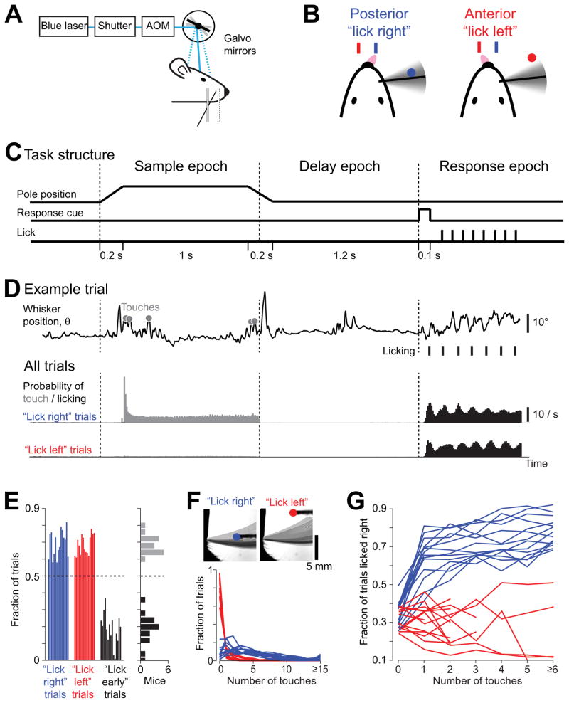
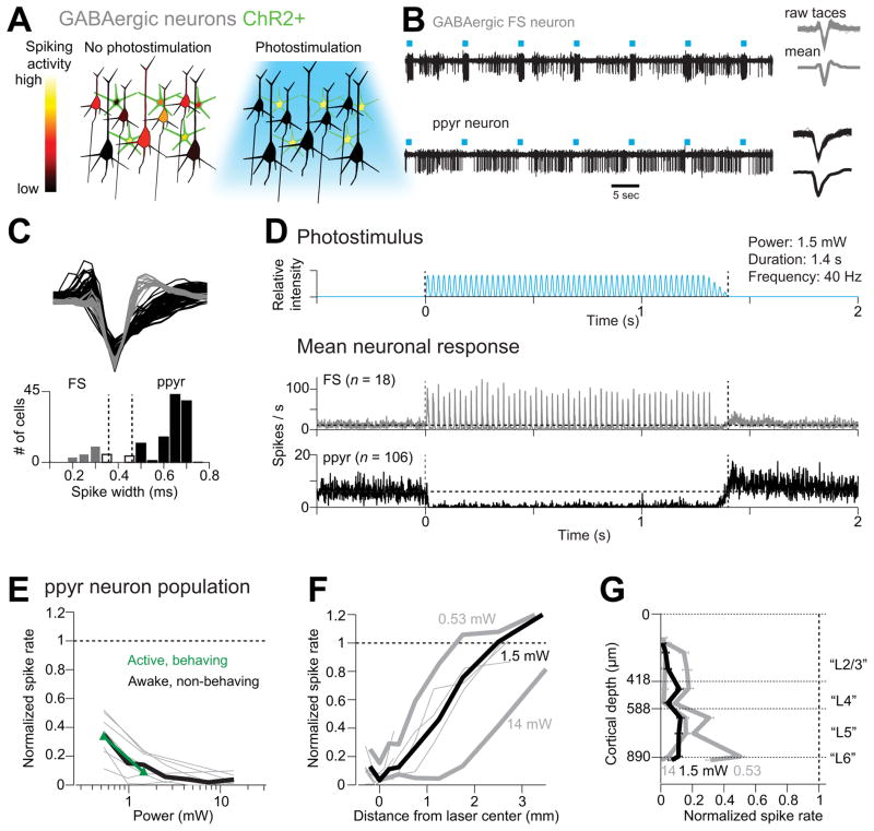
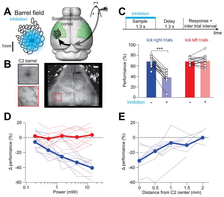
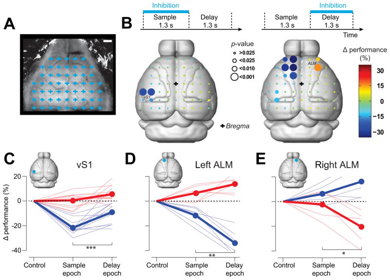
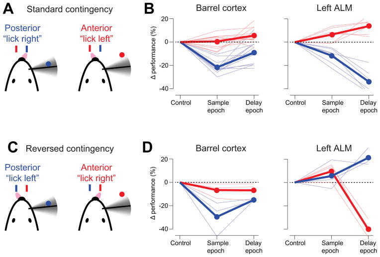
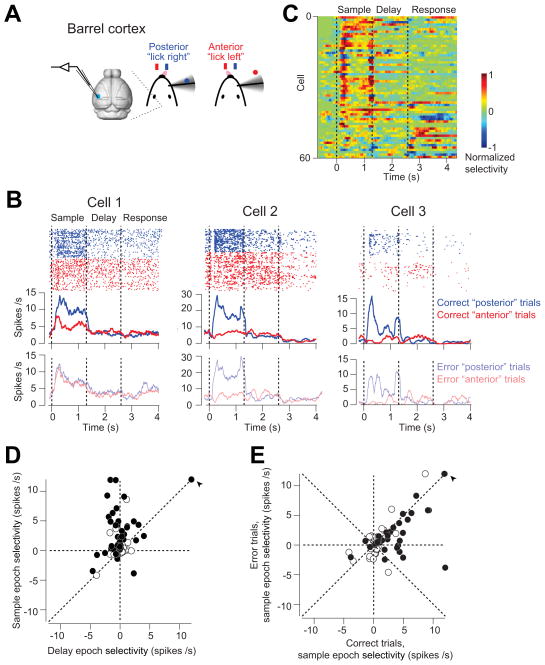
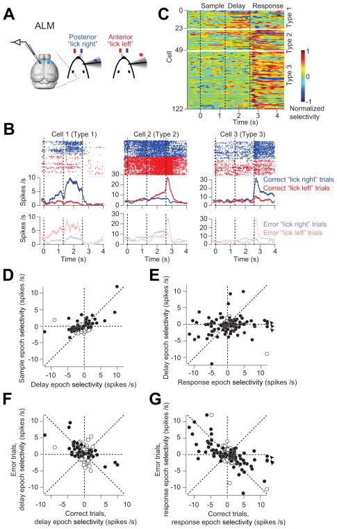
Perceptual decisions involve distributed cortical activity. Does information flow sequentially from one cortical area to another, or do networks of interconnected areas contribute at the same time? Here we delineate when and how activity in specific areas drives a whisker-based decision in mice. A short-term memory component temporally separated tactile "sensation" and "action" (licking). Using optogenetic inhibition (spatial resolution, 2 mm; temporal resolution, 100 ms), we surveyed the neocortex for regions driving behavior during specific behavioral epochs. Barrel cortex was critical for sensation. During the short-term memory, unilateral inhibition of anterior lateral motor cortex biased responses to the ipsilateral side. Consistently, barrel cortex showed stimulus-specific activity during sensation, whereas motor cortex showed choice-specific preparatory activity and movement-related activity, consistent with roles in motor planning and movement. These results suggest serial information flow from sensory to motor areas during perceptual decision making.


Conflict of interest statement

The authors declare no competing interests.

Figures

Figure 1
Figure 1. Object location discrimination task with a delay epoch
(A) Head-fixed mouse performing object location discrimination under optogenetic perturbation. (B) A mouse producing “lick right” and “lick left” responses based on pole location. (C) Task structure. The pole was within reach during the sample epoch. Mice responded with licking after an auditory response cue. (D) Behavioral data. Top, one example trial. Whisker position (azimuthal angle, θ) for a representative “lick right” trial. Touches, grey circles; licks, black ticks. Middle, Summary data for “lick right” trials in 8 mice. Probability of touch, grey; licking, black (10 ms time bin). Bottom, same as middle for “lick left” trials. (E) Behavioral performance across mice. Left, fraction correct “lick right” (blue), “lick left” (red), and “lick early” (black) trials. Each bar corresponds to one mouse (n = 15). Right panel, histogram of performance (grey) and fraction of “lick early” trials (black) across individual mice. (F) Top, whisker density for representative “lick right” and “lick left” trials (overlaid whisker images). Bottom, distribution of the number of touches in “lick right” and “lick left” trials. Each line corresponds to one mouse (n = 14). (G)The fraction of “lick right” responses as a function of number of touches for “lick right” (blue) and “lick left” (red) trials (n = 14). One mouse from (E) was excluded because there were not enough trials to sort by the number of touches (Table 1). See also Figure S1.
Figure 2
Figure 2. ChR2-assisted photoinhibition
(A) Inactivation by photostimulating ChR2-positive GABAergic interneurons (green). (B) Silicon probe recordings. Top, a GABAergic fast spiking (FS) neuron (other units with smaller spike amplitudes were also recorded on this electrode). Bottom, a putative pyramidal (ppyr) neuron. Right, corresponding spike waveforms. (C) Spike classification. Top, spike waveforms for FS neurons (n = 18; grey) and ppyr neurons (n = 106; black). Bottom, histogram of spike durations. Neurons that could not be classified based on spike width were excluded from analysis (white bar, n = 9; see Experimental Procedures). (D) Top, the photostimulus. Vertical dotted lines, start and stop of photostimulation. Bottom, mean peristimulus time histogram (PSTH, 1 ms bin) for FS neurons and ppyr neurons recorded under awake, non-behaving conditions. All neurons <0.25 mm from the laser center were pooled. (E) Spike rate as a function of laser power (<1 mm from laser center, all cortical depths). Spike rates were normalized to baseline (dash line, see Experimental Procedures). Thick black line, mean for awake, non-behaving condition. Thin grey lines, individual mice (7 mice, 103 ppyr neurons; one mouse with only 3 ppyr neurons was excluded). Green line, mean for active behaving condition (35 neurons, 6 mice; error bars reflect s.e.m. over mice). (F) Normalized spike rate versus distance from the photostimulus center (all cortical depths). Neurons were pooled across cortical depths. Thin lines, individual mice for the 1.5 mW condition. (G)Normalized spike rate versus cortical depth (< 0.2 mm from laser center). Recording depths and cortical layers (“L”) are based on histology. Error bars indicate s.e.m. over neurons (n = 106). See also Figures S2, S3, S4.
Figure 3
Figure 3. Photoinhibition of vS1 during object location discrimination
(A) Approximate spatial extent of photoinhitibion under our standard condition (1.5 mW). Photoinhibition spans at least 10 barrel columns. Right, the primary somatosensory cortex (green), with the barrel field superposed. (B) Mapping the C2 column with iIntrinsic signal imaging (top left) relative to vasculature landmarks (bottom left). Right, an example clear-skull cap. Scale bar, 1 mm. (C) Photoinhibition of vS1 during the sample epoch. Top, time-line of photoinhibition. Bottom, effects of photoinhibition on behavior in “lick right” trials (blue) and “lick left” trials (red). Performance is the fraction of correct reports for each trial type (Experimental procedures). Thin lines, individual mice (n = 15). Data from different laser powers are pooled (range, 0.97 to 14 mW; mean, 3.94 mW). ***, p < 0.001, two-tailed t-test. (D) Change in performance caused by photoinhibition versus laser power. Blue, “lick right” trials; red, “lick left” trials. Thick lines, mean performance; thin lines, individual mice (n = 10). (E) Change in performance in “lick right” trials versus photostimulus location from C2 barrel (n = 8). Laser power, 1.5 mW. See also Figure S5.
Figure 4
Figure 4. Cortical areas involved in object location discrimination revealed by photoinhibition
(A) A grid of 55 photostimulus locations through the clear-skull cap (grid spacing, 1 mm). Each grid location was chosen randomly for photostimulation during either sample or delay epoch (see Experimental Procedures). Scale bar, 1 mm. (B) Photoinhibition during different behavioral epochs. Top, photoinhibition during sample (left) and delay (right) epochs. Bottom, cortical regions involved in object location discrimination during sample (left) and delay (right) epochs in “lick right” trials. Color codes for the change in performance (%) under photoinhibition relative to control performance. Circle size codes for significance obtained from bootstrap (Experimental Procedures; from small to large; > 0.025, < 0.025, < 0.01, < 0.001). Effects on “lick left” trials are shown in Figure S6. Boundaries of cortical areas are from Allen Brain Atlas (Brain Expolorer 2, www.brain-map.org). (C) Photoinhibition of vS1 during the sample epoch caused a larger behavioral deficit than during the delay epoch in “lick right” trials (***, blue, “lick right” trials, p < 0.001, n = 12, two tailed t-test). Thick lines, mean; thin lines, individual mice. (D) Photoinhibition of the left ALM during the delay epoch caused a larger behavioral deficit than during the sample epoch in “lick right” trials (**, blue, “lick right” trials, p = 0.0016, n = 6; two tailed t-test; red, “no” trials, p = 0.09). (E) Photoinhibition of the right ALM during the delay epoch caused a larger behavioral deficit than during the sample epoch in “no” trials (blue, “lick right” trials, p = 0.22, n = 5; *, red, “no” trials, p = 0.05, two tailed t-test). See also Figure S6.
Figure 5
Figure 5. vS1 and ALM contribute differently to behavior
(A) “Lick left/lick right” task with standard contingency, where mice learned to associate posterior pole position with licking right. (B) Photoinhibition of vS1 and left ALM during the sample and delay epochs. Same as Figure 4C, D. (C) “Lick left/lick right” task with reversed contingency, where mice learned to associate posterior pole position with licking left. (D) Photoinhibition of vS1 and left ALM during the sample and delay epochs under the reversed contingency.
Figure 6
Figure 6. vS1 neurons show stimulus-specific activity
(A) vS1 recording during behavior. (B) Three example vS1 neurons during object location discrimination. Top, spike raster and PSTH for correct “posterior” (blue) and “anterior” (red) trials. Bottom, PSTH for error trials (transparent color). Averaging window, 200 ms. Dashed lines delineate behavioral epochs. (C) vS1 population selectivity. Selectivity is the difference in spike rate between the “posterior” and “anterior” trials, normalized to the peak. Averaging window, 200 ms. 15/75 vS1 neurons did not show significant selectivity during any behavioral epoch, and they were excluded from the plot. (D) vS1 neurons are mainly selective during the sample epoch. Circles correspond to individual neurons (n = 75). Selectivity is the firing rate (FR) difference between “posterior” and “anterior” trials during sample or delay epoch (FR “posterior” − FR “anterior”). Filled circles indicate neurons with significant selectivity during either the sample or delay epoch (p < 0.05, two-tailed t-test). Arrow, values are off scale; sample epoch selectivity, 19.4 spikes/s; delay epoch selectivity, 19 spikes/s. (E) vS1 maintains selectivity on error trials. Selectivity on correct trials versus error trials, slope = 0.47, r = 0.54, p < 0.001. Filled circles indicate neurons with significant sample epoch selectivity on the correct trials (p < 0.05, two-tailed t-test). Arrow, the same outlier neuron as in (C). See also Figure S7.
Figure 7
Figure 7. ALM neurons show choice-specific preparatory and movement-related activity
(A) ALM recording during behavior. (B) Three example ALM neurons during object location discrimination. Top, spike raster and PSTH for correct “lick right” (blue) and “lick left” (red) trials. Bottom, PSTH for error trials (transparent color). Averaging window, 200 ms. Dashed lines delineate behavioral epochs. (C) ALM population selectivity. Selectivity is the difference in spike rate between the preferred and non-preferred trial-type, normalized to the peak. For each neuron, we defined its preferred trial-type (“lick right” or “lick left”) using spike counts from a subset of the trials (10 trials), and the remaining data was used to compute the selectivity. Averaging window, 200 ms. Six neurons of type 1 showed significant selectivity only during the sample epoch, thus only 43 neurons showed significant delay epoch selectivity. (D) ALM neurons show choice-specific preparatory activity during the delay epoch. Selectivity is the firing rate (FR) difference between “lick right” and “lick left” trials during sample or delay epoch (FR”lick right” − FR”lick left”). Circles correspond to individual neurons (n = 186). Filled circles indicate neurons with significant selectivity during either the sample or delay epoch (p < 0.05, two-tailed t-test). Data from both left ALM and right ALM are shown. (E) ALM neurons show movement-related selectivity during the response epoch. Circles correspond to individual neurons (n = 186). Filled circles indicate neurons with significant selectivity during either the delay or response epoch (p < 0.05, two-tailed t-test). Arrows, values are off scale; response epoch selectivity, 12.5 spikes/s; 12.2 spikes/s, 16 spikes/s. (F) ALM preparatory activity during the delay epoch correlated with the animals’ behavioral choice. Selectivity on correct trials versus error trials, slope = −0.41, r = −0.46, p < 0.001. Filled circles indicate neurons with significant delay epoch selectivity on the correct trials (p < 0.05, two-tailed t-test). (G)ALM motor-related activity during the response epoch correlated with the animals’ behavioral choice. Selectivity on correct trials versus error trials, slope = −0.48, r = −0.62, p < 0.001. Filled circles indicate neurons with significant response epoch selectivity on the correct trials (p < 0.05, two-tailed t-test). Arrow, the same outlier neuron as in (D).

Comment in

References

    1. Afraz SR, Kiani R, Esteky H. Microstimulation of inferotemporal cortex influences face categorization. Nature. 2006;442:692–695. - PubMed
    1. Aravanis AM, Wang LP, Zhang F, Meltzer LA, Mogri MZ, Schneider MB, Deisseroth K. An optical neural interface: in vivo control of rodent motor cortex with integrated fiberoptic and optogenetic technology. J Neural Eng. 2007;4:S143–156. - PubMed
    1. Armstrong-James M, Fox K, Das-Gupta A. Flow of excitation within rat barrel cortex on striking a single vibrissa. J Neurosci. 1992;68:1345–1354. - PubMed
    1. Benjamini Y, Hochberg Y. Controlling the false discovery rate: a practical and powerful approach to multiple testing. Journal of the Royal Statistical Society, series B. 1995;57:289–300.
    1. Bisley JW, Zaksas D, Pasternak T. Microstimulation of cortical area MT affects performance on a visual working memory task. Journal of neurophysiology. 2001;85:187–196. - PubMed

Publication types

MeSH terms

Substances