. 2014 Jan 16;156(1-2):158-69.
doi: 10.1016/j.cell.2013.11.031. Epub 2013 Dec 19.

The N-terminal methionine of cellular proteins as a degradation signal

Affiliations

The N-terminal methionine of cellular proteins as a degradation signal

Heon-Ki Kim et al. Cell. .

Abstract

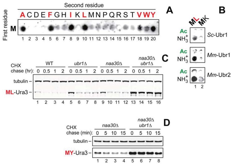
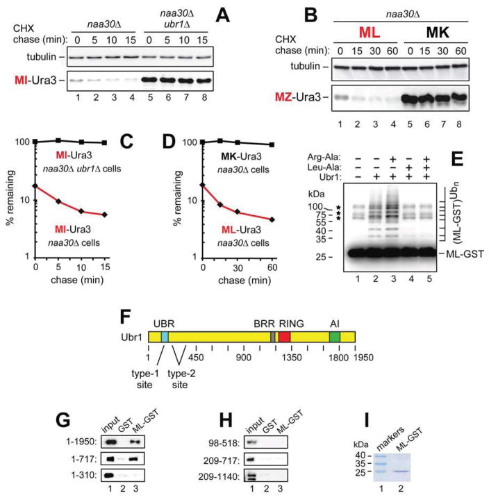
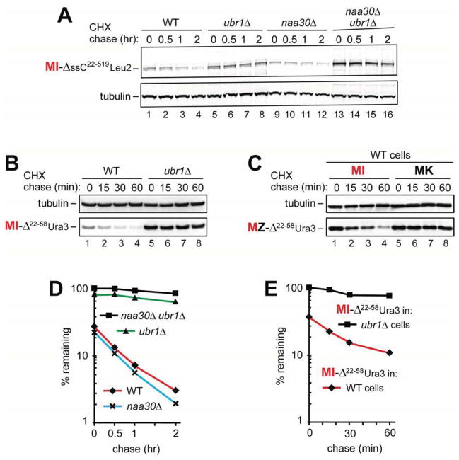
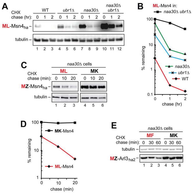
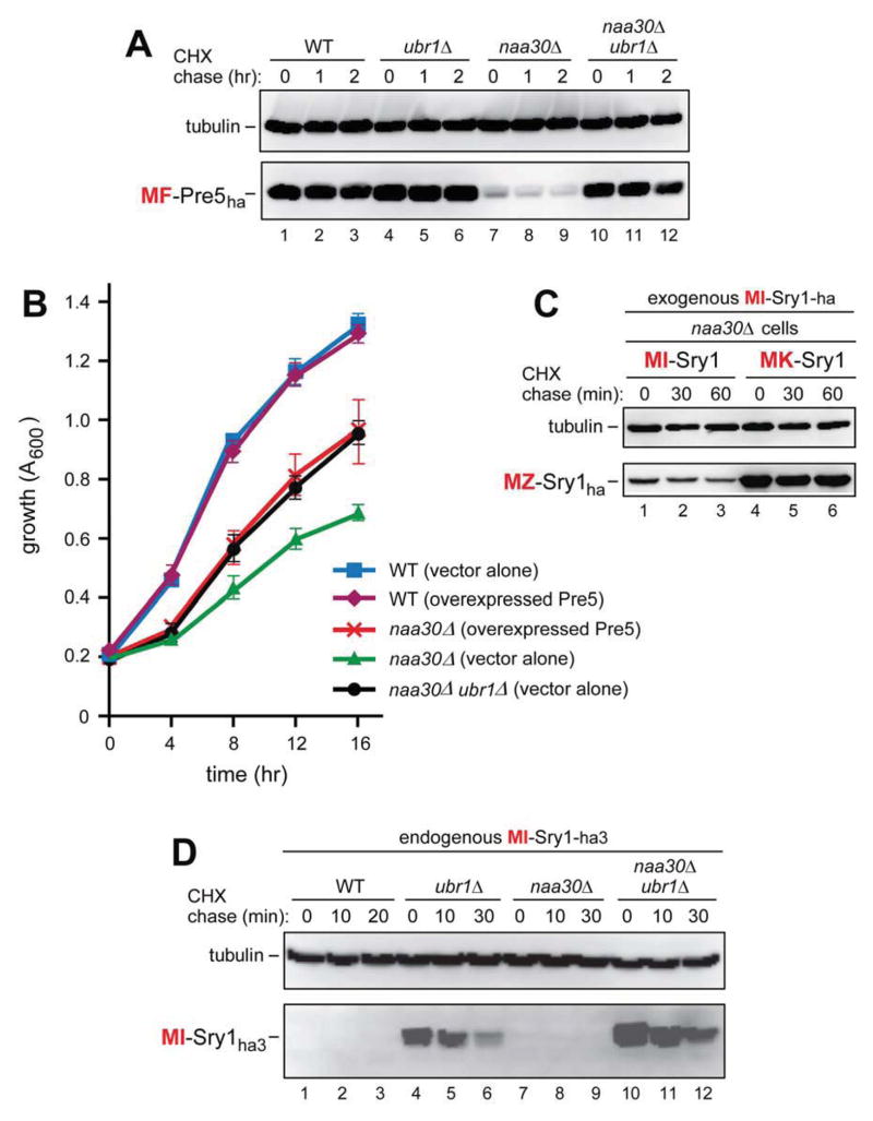
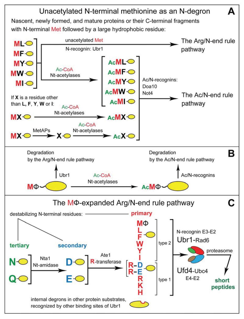
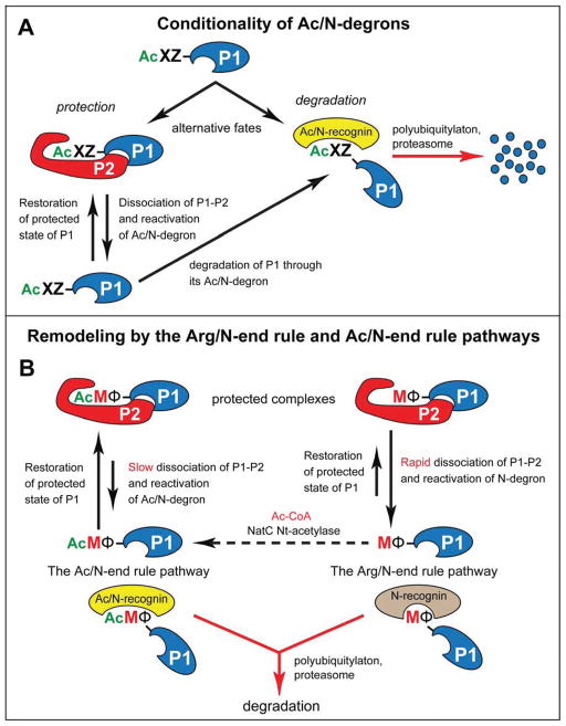
The Arg/N-end rule pathway targets for degradation proteins that bear specific unacetylated N-terminal residues while the Ac/N-end rule pathway targets proteins through their N(α)-terminally acetylated (Nt-acetylated) residues. Here, we show that Ubr1, the ubiquitin ligase of the Arg/N-end rule pathway, recognizes unacetylated N-terminal methionine if it is followed by a hydrophobic residue. This capability of Ubr1 expands the range of substrates that can be targeted for degradation by the Arg/N-end rule pathway because virtually all nascent cellular proteins bear N-terminal methionine. We identified Msn4, Sry1, Arl3, and Pre5 as examples of normal or misfolded proteins that can be destroyed through the recognition of their unacetylated N-terminal methionine. Inasmuch as proteins bearing the Nt-acetylated N-terminal methionine residue are substrates of the Ac/N-end rule pathway, the resulting complementarity of the Arg/N-end rule and Ac/N-end rule pathways enables the elimination of protein substrates regardless of acetylation state of N-terminal methionine in these substrates.


Figures

Figure 1
Figure 1. Specific Binding of Ubr1 to Unacetylated N-Terminal Methionine Followed by a Hydrophobic Residue
(A) SPOT assay with S. cerevisiae fUbr1 and Met-Z-eK(3–10) peptides MZGSGAWLLP (Z = Ala, Cys, Asp, Glu, Phe, Gly, His, Ile, Lys, Leu, Met, Asn, Pro, Gln, Arg, Ser, Thr, Val, Trp, Tyr). (B) Same as in A but with S. cerevisiae fUbr1 (ScfUbr1), mouse fUbr1 (MmfUbr1) and mouse fUbr2 (MmfUbr2) vs. Met-Z-eK(3–10) (Z = Leu, Lys) peptides and their Nt-acetylated counterparts. (C) Cycloheximide (CHX) chases with ML-Ura3 in WT (lanes 1–4), ubr1Δ (lanes 5–8), naa30Δ (lanes 9–12), and naa30Δ ubr1Δ cells (lanes 13–16). (D) CHX chases with MY-Ura3 in naa30Δ (lanes 1–4) and naa30Δ ubr1Δ cells (lanes 5–8). See also Figures S1–S3.
Figure 2
Figure 2. Unacetylated N-Terminal Methionine as an N-Degron of the Arg/N-End Rule Pathway
(A) CHX chases with MI-Ura3 in naa30Δ (lanes 1–4) and naa30Δ ubr1Δ cells (lanes 5–8). (B) CHX chases with ML-Ura3 (lanes 1–4) and MK-Ura3 (lanes 5–8) in naa30Δ cells. (C) Quantification of data in A. (D) Quantification of data in B. In A–D, the corresponding CHX-chase assays were carried out at least three times and yielded results within 10% of the data shown. (E) In vitro polyubiquitylation of purified ML-GST by the purified Ubr1-Rad6 Ub ligase. Lane 1, complete assay but without Ubr1 (the asterisks indicate three minor contaminants in purified ML-GST, whose band is indicated on the right). Lane 2, same as lane 1 but with Ubr1. Lane 3, same as lane 2 but with Arg-Ala (1 mM). Lane 4, same as lane 2 but with Leu-Ala (1 mM). Lane 5, same as lane 2 but with Arg-Ala and Leu-Ala. (F) The UBR, BRR, RING, and AI regions of the S. cerevisiae Ubr1 N-recognin (Varshavsky, 2011). (G) GST-pulldown assays with ML-GST versus GST and either full-length fUbr11–1950 or its fragments fUBR1–717 and fUbr11–310. (H) Same as in G but with fUbr198–518, fUbr1209–717 and Ubr1209–1140f. (I) SDS-PAGE of purified ML-GST (lane 2). See also Figures S1–S4.
Figure 3
Figure 3. Misfolded Proteins Containing Met-Based N-Degrons
(A) CHX chases with MI-ΔssC22–519Leu2myc in WT (lanes 1–4), ubr1Δ (lanes 5–8), naa30Δ (lanes 9–12), and naa30Δ ubr1Δ cells (lanes 13–16). (B) CHX-chases with MI-ΔssC22–58Ura3ha in WT (lanes 1–4) and ubr1Δ cells (lanes 5–8). (C) CHX-chases with MI-ΔssC22–58 Ura3ha (lanes 1–4) and MK-ΔssC22–58 Ura3ha (lanes 5–8) in WT cells. (D) Quantification of data in A. (E) Quantification of data in B. In A–E, the corresponding CHX-chase assays were carried out at least three times and yielded results within 10% of the data shown. See also Figures S1 and S2.
Figure 4
Figure 4. The Natural ML-Msn4 and MF-Arl3 Proteins Contain Met-Based N-Degrons
(A) CHX chases with ML-Msn4ha in WT (lanes 1–3), ubr1Δ (lanes 4–6), naa30Δ (lanes 7–9), and naa30Δ ubr1Δ cells (lanes 10–12). (B) Quantification of data in A. (C) CHX chases with ML-Msn4ha (lanes 1–3) and MK-Msn4ha (lanes 4–6) in naa30Δ cells. (D) Quantification of data in C. (E) CHX chases with MF-Arl3ha2 (lanes 1–3) and MK-Arl3ha2 (lanes 4–6) in naa30Δ cells. In A–E, the corresponding CHX-chase assays were carried out at least three times and yielded results within 10% of the data shown. See also Figures S1 and S2.
Figure 5
Figure 5. The Natural MF-Pre5 and MI-Sry1 Proteins Contain Met-Based N-Degrons
(A) CHX chases with MF-Pre5ha in WT (lanes 1–3), ubr1Δ (lanes 4–6), naa30Δ (lanes 7–9), and naa30Δ ubr1Δ cells (lanes 10–12). (B) Growth rates of WT, naa30Δ and naa30Δ ubr1Δ S. cerevisiae strains, including WT and naa30Δ strains that overexpressed the MF-Pre5 proteasomal subunit. Standard errors (of triplicate measurements) are shown as well. (C) CHX chases with MI-Sry1ha (lanes 1–3) and MK-Sry1ha (lanes 4–6) in naa30Δ cells. (D) CHX chases with endogenously expressed MI-Sry1ha in WT (lanes 1–3), ubr1Δ (lanes 4–6), naa30Δ (lanes 7–9), and naa30Δ ubr1Δ cells (lanes 10–12). See also Figures S1, S2, and S4.
Figure 6
Figure 6. Complementary Specificities of the Arg/N-End Rule Pathway and the Ac/N-End Rule Pathway
(A) This diagram summarizes the main discovery of the present work, a functional complementarity between the Arg/N-end rule and Ac/N-end rule pathways. This complementarity stems from the recognition of the previously unknown MetΦ/N-degrons in Met-Φ proteins vs. the recognition of the previously characterized AcMetΦ/N-degrons in AcMet-Φ proteins. AcMetΦ/N-degrons are a subset of Ac/N-degrons in Nt-acetylated cellular proteins (Hwang et al., 2010b; Shemorry et al., 2013). Met-Φ proteins are defined, in this study, as those that bear N-terminal Met followed by a large hydrophobic (Φ) non-Met residue. (B) Condensed summary of the dual-pathway circuit shown in A. (C) The MΦ-based expansion of the Arg/N-end rule pathway in the present work, through the addition of a large set of new substrates, Met-Φ proteins. See also Figure S1.
Figure 7
Figure 7. Conditionality of Ac/N-degrons and Protein Remodeling by the N-End Rule Pathway
(A) Conditionality of Ac/N-degrons. This diagram summarizes the previously attained understanding of the dynamics of Nt-acetylated proteins vis-à-vis the Ac/N-end rule pathway (see Discussion), in conjunction with the initial discovery of the Ac/N-end rule pathway (Hwang et al., 2010b; Shemorry et al., 2013). (B) The recognition of unacetylated Met-Φ proteins by the Arg/N-end rule pathway and of Nt-acetylated AcMet-Φ proteins by the Ac/N-end rule pathway underlies the proposed remodeling of protein complexes. Owing to different rates of dissociation of Nt-acetylated (AcMet-Φ) vs. unacetylated (Met-Φ) complexes (see Discussion), the subunit-selective degradation of the unacetylated P1 (Met-Φ) subunit of a P1–P2 complex upon its dissociation would allow the replacement-mediated conversion of P1–P2 into a similar but more stable complex containing the Nt-acetylated (AcMet-Φ) counterpart of the Met-Φ P1 subunit. To maximize generality of this description, the Nt-acetylation state of the P2 protein subunit was left unspecified, in contrast to the P1 subunit. Nt-acetylation of cellular proteins is largely cotranslational. The dashed arrow signifies the current uncertainty about rates of posttranslational Nt-acetylation. See Discussion for specific ramifications of this model. See also Figure S1.

Comment in

Similar articles

Cited by

References

    1. Arnesen T, Van Damme P, Polevoda B, Helsens K, Evjenth R, Colaert N, Varhaug JE, Vandekerckhove J, Lillehaug JR, Sherman F, et al. Proteomics analyses reveal the evolutionary conservation and divergence of N-terminal acetyltransferases from yeast to humans. Proc Natl Acad Sci USA. 2009;106:8157–8162. - PMC - PubMed
    1. Bachmair A, Finley D, Varshavsky A. In vivo half-life of a protein is a function of its amino-terminal residue. Science. 1986;234:179–186. - PubMed
    1. Behnia R, Panic B, Whyte JRC, Munro S. Targeting of the Arf-like GTPase Arl3p to the Golgi requires N-terminal acetylation and the membrane protein Sys1p. Nat Cell Biol. 2004;6:405–413. - PubMed
    1. Brower CS, Piatkov KI, Varshavsky A. Neurodegeneration-associated protein fragments as short-lived substrates of the N-end rule pathway. Mol Cell. 2013;50:161–171. - PMC - PubMed
    1. Choi WS, Jeong BC, Joo YJ, Lee MR, Kim J, Eck MJ, Song HK. Structural basis for the recognition of N-end rule substrates by the UBR box of ubiquitin ligases. Nat Struct Mol Biol. 2010;17:1175–1181. - PubMed

Publication types