. 2014 Apr 3;426(7):1483-97.
doi: 10.1016/j.jmb.2013.12.015. Epub 2013 Dec 17.

The solution structure of the regulatory domain of tyrosine hydroxylase

Affiliations

The solution structure of the regulatory domain of tyrosine hydroxylase

Shengnan Zhang et al. J Mol Biol. .

Abstract

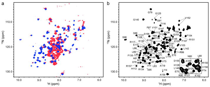
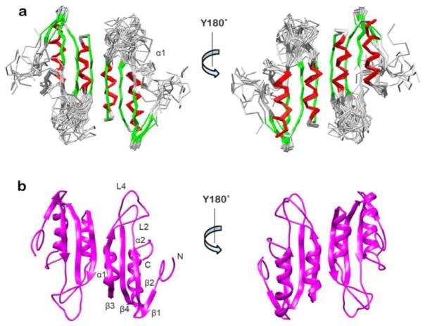
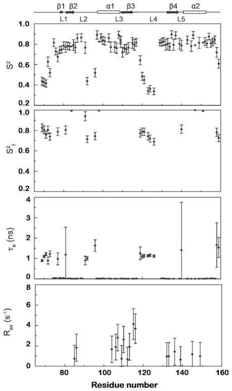
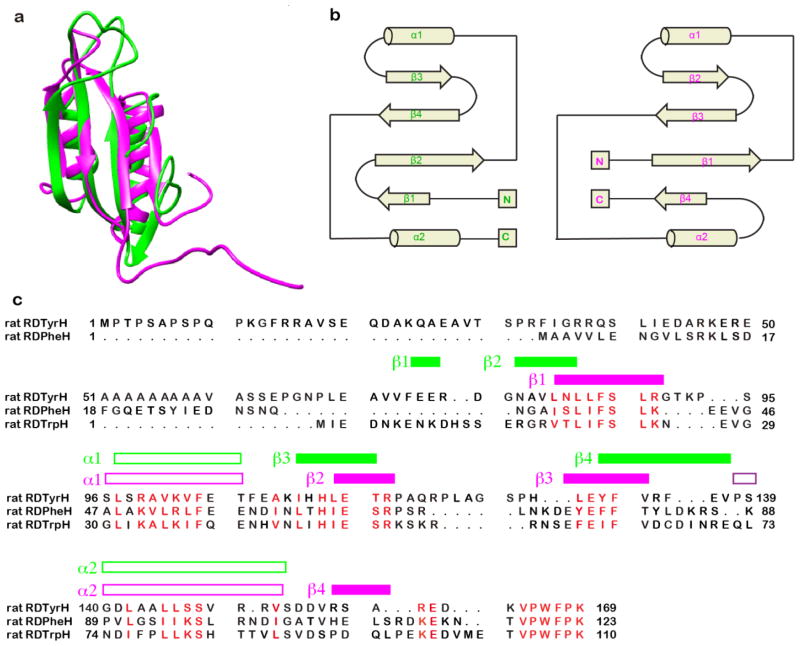
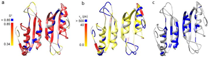
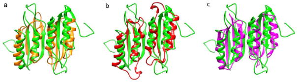
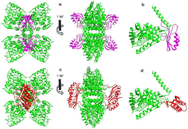
Tyrosine hydroxylase (TyrH) catalyzes the hydroxylation of tyrosine to form 3,4-dihydroxyphenylalanine in the biosynthesis of the catecholamine neurotransmitters. The activity of the enzyme is regulated by phosphorylation of serine residues in a regulatory domain and by binding of catecholamines to the active site. Available structures of TyrH lack the regulatory domain, limiting the understanding of the effect of regulation on structure. We report the use of NMR spectroscopy to analyze the solution structure of the isolated regulatory domain of rat TyrH. The protein is composed of a largely unstructured N-terminal region (residues 1-71) and a well-folded C-terminal portion (residues 72-159). The structure of a truncated version of the regulatory domain containing residues 65-159 has been determined and establishes that it is an ACT domain. The isolated domain is a homodimer in solution, with the structure of each monomer very similar to that of the core of the regulatory domain of phenylalanine hydroxylase. Two TyrH regulatory domain monomers form an ACT domain dimer composed of a sheet of eight strands with four α-helices on one side of the sheet. Backbone dynamic analyses were carried out to characterize the conformational flexibility of TyrH65-159. The results provide molecular details critical for understanding the regulatory mechanism of TyrH.

Keywords: ACT domain; NMR spectroscopy; regulation; solution structure; tyrosine hydroxylase.


Figures

Figure 1
Figure 1
NMR spectra of the isolated regulatory domain of tyrosine hydroxylase. (a) Overlay of the 2D 1H-15N HSQC spectra of RDTyrH (red) and RDTyrH65–159 (blue). (b) 2D 1H-15N HSQC spectrum of 0.8 mM RDTyrH65–159 showing the assignments of the individual residues. Conditions: 50 mM sodium phosphate, 1 μM leupeptin, 1 μM pepstatin A and 5% D2O (pH 7.0), at 300 K at a magnetic field strength of 14.1 T (600 MHz 1H).
Figure 2
Figure 2
Structure of the regulatory domain of tyrosine hydroxylase. (a) Superposition of the backbones of the 10 lowest-energy structures of RDTyrH65–159 in two orientations with an 180° rotation along the Y-axis. β-Strands are in green and helices in red. (b) Ribbon diagram of a representative low-energy structure in two orientations with an 180° rotation along the Y-axis indicating the location of secondary structure elements.
Figure 3
Figure 3
Model-free parameters for RDTyrH65–159 backbone amides derived by the fitting of the 15N T1, 15N T2, and 1H-15N NOE data. Lipari-Szabo S2, S2f, τe, and Rex parameters are shown from top to bottom, respectively. Missing S2, S2f, τe, and Rex data points indicate that this parameter was not included in the motional model for that residue. A representation of the RDTyrH65–159 secondary structure is shown along the top.
Figure 4
Figure 4
Comparison of the structures of the regulatory domains of phenylalanine hydroxylase and tyrosine hydroxylase. (a) Overlay of the ribbon diagrams of RDTyrH65–159 (green) and RDPheH (medium purple, residues 19–117, PDB code 2PHM). (b) Topology diagrams of RDTyrH65–159 (top) and RDPheH (bottom); the structural elements are labeled in green and magenta, respectively. (c) Structure-based sequence alignment of the regulatory domains of the three amino acid hydroxylases. The secondary structural elements of RDTyrH65–159 and RDPheH are labeled green and magenta, respectively. To generate the alignment, a structure-based alignment of RDTyrH65–159 and RDPheH was first performed with Chimera. The sequence of the regulatory domain of TrpH was then added to the alignment using Clustal X.
Figure 5
Figure 5
The dynamic properties of the backbone of the RDTyrH65–159 dimer: a) the generalized order parameter S2 values with colors ranging from yellow to red and blue corresponding to S2 values from 0.4 to 0.85, and >0.85; b) the internal motions on the ps to ns time scales with colors ranging from yellow to red and magenta corresponding to Te values from 0 to 40 ps and >500 ps; c) residues with conformational changes (Rex) on the microsecond to millisecond time scales colored in blue. Residues for which the respective parameter was not included in the model are colored in grey.
Figure 6
Figure 6
Structural comparisons between the RDTyrH65–159 dimer (green) and three ACT domain dimers: (a) phosphoglycerate dehydrogenase (orange, PDB code 1PSD), (b) aspartate kinase from Arabidopsis (red, PDB code 2CDQ), and (c) E. coli IlvH, the regulatory subunit of acetohydroxyacid synthase (magenta, PDB code 2F1F).
Figure 7
Figure 7
Structural models of the combined regulatory, catalytic, and tetramerization domains of TyrH. a, b: Models of the tetramer (a) and monomer (b) derived from the structure of PheH (PDB code 2PHM) by replacing the regulatory domain of PheH with the RDTyrH65–159 monomer (magenta) and the catalytic domain of PheH with the combined catalytic and tetramerization domains of TyrH (PDB code 1TOH, green), respectively. c, d: Models of the tetramer (c) and monomer (d) derived from the model of Jaffe et al. by replacing the dimer of the regulatory domain of PheH with the RDTyrH65–159 dimer (red) and the catalytic and tetramerization domains of PheH with the catalytic and tetramerization domains of TyrH (green), respectively. The missing residues connecting the two domains were added with Modeller in Chimera.

References

    1. Rao F, Zhang K, Zhang L, Rana B, Wessel J, Fung M, Rodriguez-Flores J, Taupenot L, Ziegler M, O’Connor D. Human tyrosine hydroxylase natural allelic variation: Influence on autonomic function and hypertension. Cell Mol Neurobiol. 2010;30:1391–1394. - PMC - PubMed
    1. Hoffmann GF, Assmann B, Brautigam C, Dionisi-Vici C, Haussler M, de Klerk JB, Naumann M, Steenbergen-Spanjers GC, Strassburg HM, Wevers RA. Tyrosine hydroxylase deficiency causes progressive encephalopathy and dopa-nonresponsive dystonia. Ann Neurol. 2003;54(Suppl 6):S56–S65. - PubMed
    1. Ludecke B, Dworniczak B, Bartholome K. A point mutation in the tyrosine hydroxylase gene associated with Segawa’s syndrome. Hum Genet. 1995;95:123–125. - PubMed
    1. Fitzpatrick PF. Tetrahydropterin-dependent amino acid hydroxylases. Ann Rev Biochem. 1999;68:355–381. - PubMed
    1. Lohse DL, Fitzpatrick PF. Identification of the intersubunit binding region in rat tyrosine hydroxylase. Biochem Biophys Res Commun. 1993;197:1543–1548. - PubMed

Publication types

MeSH terms

Substances

LinkOut - more resources