. 2014 Apr 22;63(15):1556-66.
doi: 10.1016/j.jacc.2013.11.023. Epub 2013 Dec 18.

In vivo silencing of the transcription factor IRF5 reprograms the macrophage phenotype and improves infarct healing

Affiliations

In vivo silencing of the transcription factor IRF5 reprograms the macrophage phenotype and improves infarct healing

Gabriel Courties et al. J Am Coll Cardiol. .

Abstract

Objectives: The aim of this study was to test whether silencing of the transcription factor interferon regulatory factor 5 (IRF5) in cardiac macrophages improves infarct healing and attenuates post-myocardial infarction (MI) remodeling.

Background: In healing wounds, the M1 toward M2 macrophage phenotype transition supports resolution of inflammation and tissue repair. Persistence of inflammatory M1 macrophages may derail healing and compromise organ functions. The transcription factor IRF5 up-regulates genes associated with M1 macrophages.

Methods: Here we used nanoparticle-delivered small interfering ribonucleic acid (siRNA) to silence IRF5 in macrophages residing in MIs and in surgically-induced skin wounds in mice.

Results: Infarct macrophages expressed high levels of IRF5 during the early inflammatory wound-healing stages (day 4 after coronary ligation), whereas expression of the transcription factor decreased during the resolution of inflammation (day 8). Following in vitro screening, we identified an siRNA sequence that, when delivered by nanoparticles to wound macrophages, efficiently suppressed expression of IRF5 in vivo. Reduction of IRF5 expression, a factor that regulates macrophage polarization, reduced expression of inflammatory M1 macrophage markers, supported resolution of inflammation, accelerated cutaneous and infarct healing, and attenuated development of post-MI heart failure after coronary ligation as measured by protease targeted fluorescence molecular tomography-computed tomography imaging and cardiac magnetic resonance imaging (p < 0.05).

Conclusions: This work identified a new therapeutic avenue to augment resolution of inflammation in healing infarcts by macrophage phenotype manipulation. This therapeutic concept may be used to attenuate post-MI remodeling and heart failure.

Keywords: IRF5; healing; heart failure; macrophage; myocardial infarction.


Figures

Figure 1
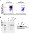
Figure 1. Upregulation of IRF5 in heart macrophages after MI
(A) Representative flow cytometric dot plots show samples from day 4 after MI, stained with lineage cocktail and CD11b antibodies before and after MACS separation. (B) Unlabeled fractions were processed for western blot analysis of IRF5 in isolated cells from naive hearts, infarct tissue and remote myocardium. Liver mouse tissue was used as a positive control.
Figure 2
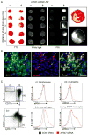
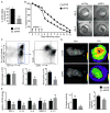
Figure 2. Infarct uptake of LNP-siRNA nanoparticles
(A) TTC staining and ex-vivo fluorescence reflectance imaging (FRI) of 4 day-old infarcts, 2 hours following i.v. injection of LNP-encapsulated AF647-labelled siRNA. (B) Immunofluorescence microscopy of infarcts showing colocalization of CD11b expressing cells (green) and fluorescently labeled siRNA (red). Scale bar indicates 50 μm. (C) FACS of siRNA uptake by lymphocytes (i), neutrophils (ii), macrophages (iv) and Ly-6Chigh monocytes (v) isolated from infarcts.
Figure 3
Figure 3. LNP-IRF5 siRNA treatment results in efficient silencing in macrophages
(A) IRF5 siRNA candidates were ordered by their silencing efficiency at 10 nM concentration. The red frame highlights the siRNA selected for subsequent in vivo experiments. (B) Mice were injected i.v. with either the LNP-IRF5 siRNA (siIRF) or control siRNA (siCON). Twenty-four hours later, splenic Ly-6Chigh monocytes were isolated by FACS to quantify IRF5 mRNA (n=7 per group). (C) IRF5 mRNA and (D) IRF5 protein levels in infarct macrophages on day 4 after MI. *P<0.05, **P<0.01, ***P<0.001.
Figure 4
Figure 4. IRF5 silencing modulates macrophage polarization in apoE−/− mice with MI
(A) FACS plots of infarct tissue and analysis of IRF5 protein in macrophages and Ly-6Chigh monocytes. Histogram plots display control siRNA (red), IRF5 siRNA (blue) and isotype control (grey). Bar graph shows mean fluorescence intensity (MFI) for IRF5 (n=8 per group). (B) IRF5 gene expression in infarct macrophages (n = 6–8 per group). (C) Neutrophils (ii), macrophages (iv) and Ly-6Chigh monocytes (v) in infarcts on day 4 after MI. (D) M1 and M2 genes in macrophages isolated from 4 day old infarcts (n = 8–11 per group). *P<0.05, **P<0.01.
Figure 5
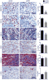
Figure 5. Histological assessment of infarct healing on day 7 after MI in ApoE−/− mice treated with siCON or siIRF5
Immunohistochemistry staining for myeloid cells (NIMP-R14; CD11b), macrophages (MAC-3), neo-vascularization (CD31), collagen deposition (collagen I) and myofibroblasts (α-SMA). Bar graphs display region of interest (ROI, n=5–7 per group). *P<0.05, **P<0.01, ***P<0.001.
Figure 6
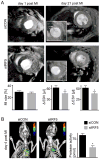
Figure 6. siIRF5 improves infarct healing in ApoE−/− mice
(A) Serial cardiac MRI (day 1 and day 21) in ApoE−/− mice treated with siIRF5 or siCON. The dotted line highlights the infarct. Insets show the infarct-induced thinning of the left ventricular wall in apical short axis slices. (B) FMT-CT on day 4 after MI. Circles indicate infarct signal (n=5–9 per group). *P<0.05.
Figure 7
Figure 7. LNP-siRNA delivery to monocytes/macrophages in skin wounds
(A) On day 4 after wound induction, mice were injected i.v. with AF647 labeled siRNA encapsulated in lipid-nanoparticles. Fluorescence reflectance imaging (FRI) revealed a strong siRNA signal in the wound one hour after injection. (B) FACS analysis of siRNA uptake by both F4/80high macrophages and Ly-6Chigh monocytes in the wound. (C) Quantitation of protease activity in skin wounds by FRI revealed the highest signal peaking on day 4 post-injury (n=6 per time point). *P <0.01.
Figure 8
Figure 8. Treatment with siIRF5 accelerates skin wound healing
(A) IRF5 silencing in monocytes/macrophages isolated from the wound (n=9 per group). (B) Wound area (n = 7–8 per group). (C) FACS of wound leukocyte content on day 4 (n=5 per group). (D) Protease activity by FMT and FRI. TBR, target to background ratio (n=8 per group). (e) M1 and M2 genes in wound macrophages on day 4 (n=10–12 per group). *P<0.05, **P<0.01, ***P<0.001.

Comment in

References

    1. Singer AJ, Clark RA. Cutaneous wound healing. N Engl J Med. 1999;341:738–46. - PubMed
    1. Mantovani A, Biswas SK, Galdiero MR, Sica A, Locati M. Macrophage plasticity and polarization in tissue repair and remodelling. J Pathol. 2013;229:176–85. - PubMed
    1. Biswas SK, Chittezhath M, Shalova IN, Lim JY. Macrophage polarization and plasticity in health and disease. Immunol Res. 2012;53:11–24. - PubMed
    1. Swirski FK, Nahrendorf M. Leukocyte behavior in atherosclerosis, myocardial infarction, and heart failure. Science. 2013;339:161–66. - PMC - PubMed
    1. Lambert JM, Lopez EF, Lindsey ML. Macrophage roles following myocardial infarction. Int J Cardiol. 2008;130:147–58. - PMC - PubMed

Publication types